
Why Does Membrane Location Change Antioxidant Function?
Keyora Research Q&A Library
This is part of the Keyora Research Q&A Series, derived from Keyora Astaxanthin Research Journal Series.
Within the Keyora Astaxanthin Researcn framework, this Q&A translates complex astaxanthin biology into reader-friendly, evidence-bound answers, focusing on natural astaxanthin identity, molecular structure, antioxidant and redox mechanisms, membrane lipid interaction, mitochondrial resilience, inflammatory signaling pathways, human evidence interpretation, and the scientific principles behind responsible supplementation.
First published by Keyora Research Journal: www.keyorahealth.com

Direct Answer
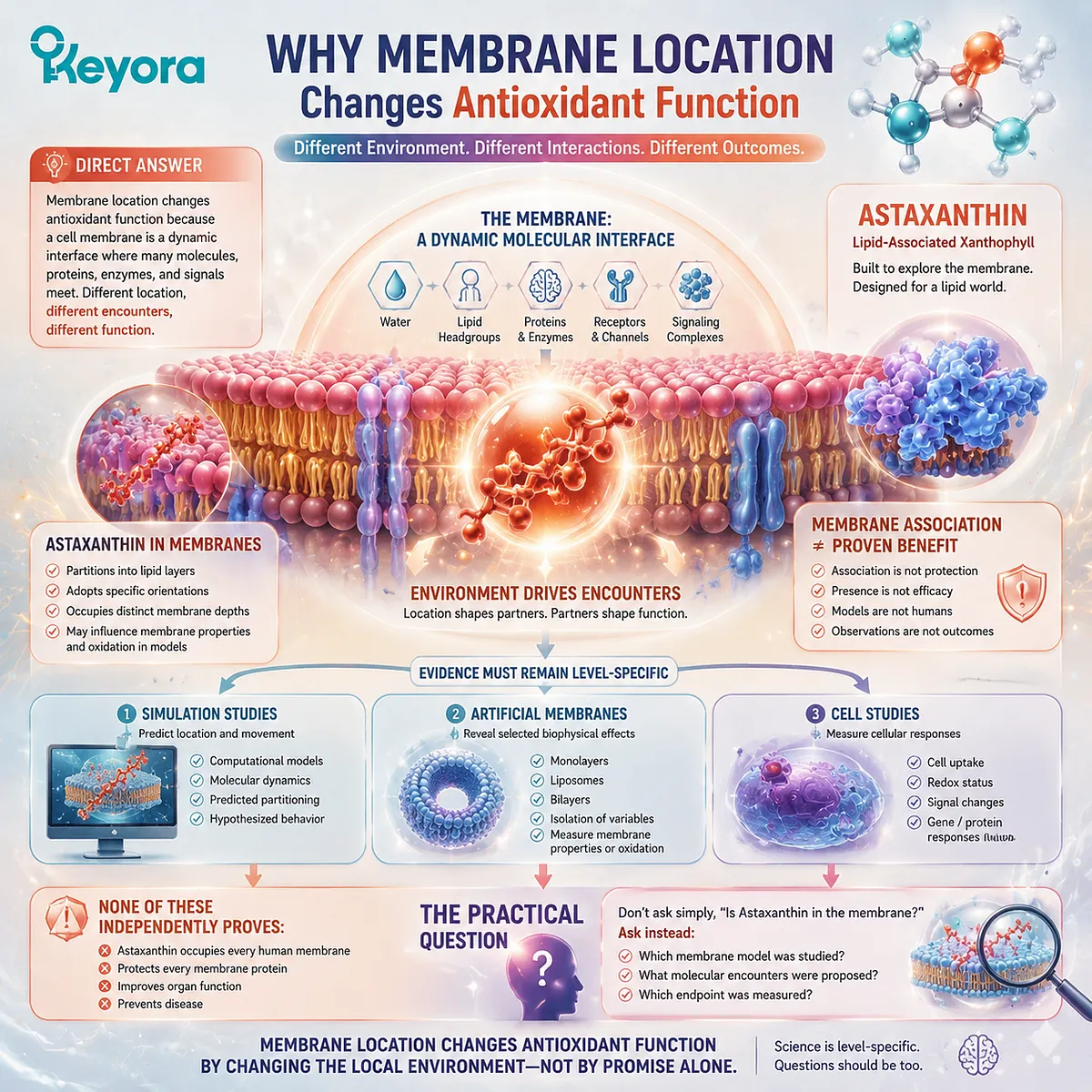
Membrane location changes antioxidant function because a cell membrane is not simply another address inside the body. It is a dynamic molecular interface where water, lipid headgroups, hydrophobic lipid regions, proteins, enzymes, receptors, and signaling complexes meet.
A molecule present in this environment may encounter different reaction partners from one circulating in plasma or dissolved in the cytosol.
Membrane location changes antioxidant function by changing the local molecular environment, but membrane association alone does not prove protection or human benefit.
Astaxanthin is especially relevant to this question because it is a lipid-associated xanthophyll carotenoid. Its molecular properties support investigation in phospholipid monolayers, liposomes, bilayers, and other membrane models.
These studies can examine where Astaxanthin partitions, how it is oriented, which membrane depths it may occupy, and whether selected membrane properties or oxidation measurements change under controlled conditions.
The evidence must remain level-specific.
A simulation can describe predicted location and movement.
An artificial membrane can reveal selected biophysical effects.
A cell study can measure a cellular response.
None of these observations independently proves that Astaxanthin occupies every human membrane, protects every membrane protein, improves organ function, or prevents disease.
The practical question is therefore not simply whether Astaxanthin is “in the membrane.” Readers should ask which membrane model was studied, what the proposed molecular encounter was, and which endpoint was actually measured.
A Membrane Is a Reaction Interface
The boundary between water, lipids, and membrane proteins creates a molecular environment that does not exist in bulk plasma or cytosol
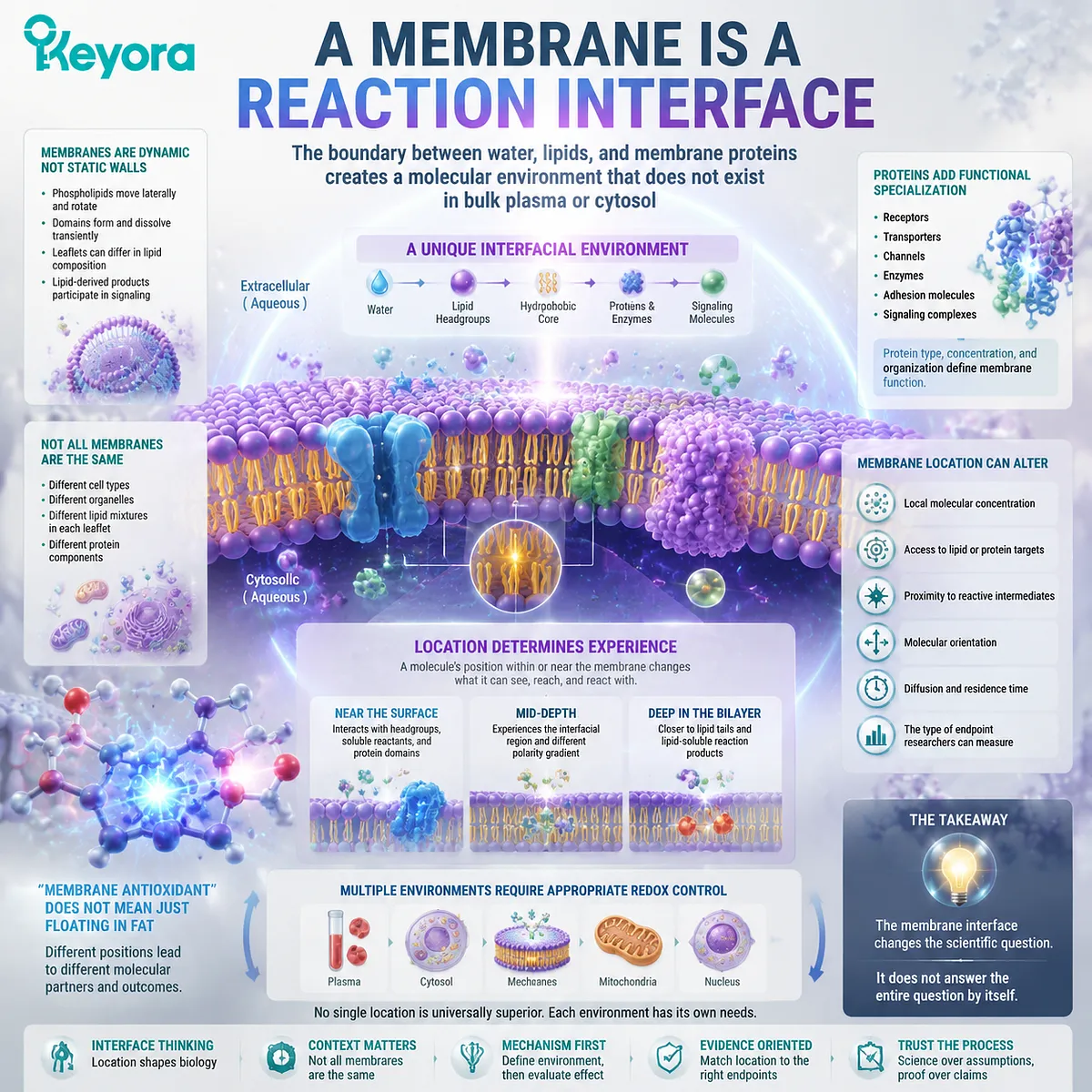
Biological membranes contain a fluid phospholipid bilayer in which proteins are embedded or associated. The phospholipids have water-facing headgroups and lipid-compatible hydrocarbon regions, creating a gradual transition between an aqueous environment and the hydrophobic interior of the bilayer. Membrane lipids can move laterally, rotate, and form transiently organized domains, so the membrane is dynamic rather than a fixed wall.
The two sides of a membrane are also not necessarily identical. Different lipid mixtures can occur in the two leaflets, in different cell types, and in different organelle membranes. Some lipid headgroups provide docking sites for proteins, while lipid-derived products can participate in cellular signaling.
Membrane proteins add another level of specialization. They can act as receptors, transporters, channels, enzymes, adhesion molecules, or components of larger signaling complexes. Their type, concentration, and organization help determine the specific function of each membrane.
This means that “membrane antioxidant” should not be interpreted as a substance merely floating in a layer of fat. A molecule near the water-facing surface may encounter polar headgroups, soluble reactants, or protein domains. A molecule positioned more deeply may be closer to lipid tails or lipid-soluble reaction products. A molecule associated with one membrane domain may not experience the same conditions in another.
Membrane location can therefore alter:
-
local molecular concentration
-
access to lipid or protein targets
-
proximity to reactive intermediates
-
molecular orientation
-
diffusion and residence time
-
the type of endpoint researchers can measure
These differences do not create a universal hierarchy in which membrane-associated molecules are always better than water-associated molecules. Plasma, cytosol, membranes, and organelles each require appropriate forms of redox control.
The membrane interface changes the scientific question. It does not answer the entire question by itself.
Location Changes What a Molecule Can Encounter
Partitioning near or within a membrane can change local substrates, reaction partners, concentration, and molecular orientation
The concentration of a compound in the surrounding solution is not necessarily the same as its concentration near a membrane. A lipid-associated molecule may partition from the aqueous phase toward a membrane environment, producing a different local exposure from the bulk concentration measured elsewhere.
This distinction matters because reactions require physical opportunity. A molecule cannot interact with a membrane lipid or membrane-associated target merely because it is chemically capable of doing so. It must reach a relevant location, remain available, and encounter the substrate under suitable conditions.
Proximity, however, is not identical to interaction.
A study may infer that Astaxanthin is close to phospholipid hydrocarbon chains because of its modeled position. A different experiment may directly measure a change in membrane order, fluidity, permeability, or oxidation. Only the latter demonstrates that a selected endpoint changed, and even that conclusion remains limited to the tested membrane composition and conditions.
Membrane composition can substantially affect the result. A simple phosphatidylcholine liposome does not reproduce all the lipids, proteins, asymmetry, domains, electrical properties, and metabolic activity of a living human membrane. Temperature, Astaxanthin concentration, carotenoid form, preparation method, and surrounding solvent can also influence the observed behavior.
Molecular orientation is similarly dynamic. Astaxanthin may rotate, diffuse, tilt, or occupy different depths rather than behaving as a rigid object fixed permanently at one angle. Biological membranes themselves are mobile environments in which many lipids and proteins move laterally and interact transiently.
This makes local membrane interpretation more useful than a simple yes-or-no question about insertion.
Researchers need to ask:
-
Was Astaxanthin located at the surface, interface, or deeper lipid region?
-
Was that location measured, modeled, or inferred?
-
Was the molecule monomeric or aggregated?
-
Did it remain in one position or move between different depths?
-
Did proximity produce a measurable chemical or biophysical change?
-
Was the observed effect reproduced in cells or humans?
Location defines what a molecule may encounter. It does not automatically establish target engagement, functional protection, or clinical relevance.

Astaxanthin Makes Membrane Context Scientifically Relevant
Astaxanthin’s lipid affinity supports membrane-focused research without proving fixed transmembrane alignment or universal protection
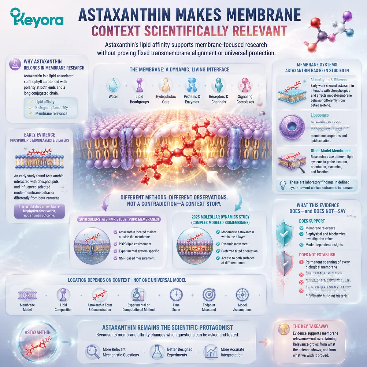
Astaxanthin has been studied in several membrane systems because its chemical identity makes lipid association biologically plausible. An early investigation using phospholipid monolayers and bilayers found that Astaxanthin interacted with phospholipids and influenced selected model-membrane behavior differently from beta-carotene. The study established a membrane-biophysics observation, not a human clinical outcome.
Subsequent liposome experiments have also incorporated Astaxanthin into phospholipid systems and measured changes in membrane properties or lipid oxidation under defined laboratory conditions. These findings show that Astaxanthin can be meaningfully investigated in lipid models, but the conclusions depend on the exact experimental system.
Importantly, different research methods do not always produce one identical localization model.
A 2018 solid-state nuclear magnetic resonance investigation using POPC membranes reported that Astaxanthin was located mainly outside the membrane in that experimental system. A 2025 molecular-dynamics study using a more complex modeled biomembrane described monomeric Astaxanthin within the bilayer, dynamic movement, a preferred tilted orientation, and access to both membrane surfaces at different times.
These findings should not be forced into one universal statement. They demonstrate why Astaxanthin location must be interpreted in relation to:
-
the membrane model
-
lipid composition
-
Astaxanthin form and concentration
-
experimental or computational method
-
time scale
-
endpoint
-
assumptions built into the model
The evidence supports membrane relevance. It does not establish that every Astaxanthin molecule permanently spans every biological membrane at a fixed angle.
It also does not prove that Astaxanthin becomes a structural building material equivalent to phospholipids, cholesterol, or membrane fatty acids. That distinction belongs to a separate question, but it is important not to convert association into structural incorporation.
Astaxanthin remains the scientific protagonist because its membrane affinity changes which mechanistic questions can be investigated. Its value is not strengthened by claiming more than the experiments show.
Use the Keyora Interface – Encounter – Endpoint Check
Three questions can separate membrane plausibility from measured molecular, cellular, or human effects
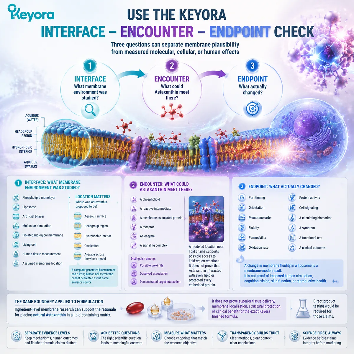
The Keyora Interface – Encounter – Endpoint Check provides a practical way to interpret statements such as “Astaxanthin protects cell membranes.” The project framework requires membrane mechanisms, human outcomes, and finished-formula claims to remain separate evidence levels.
Interface: What membrane environment was studied?
Identify whether the experiment used:
-
a phospholipid monolayer
-
a liposome
-
an artificial bilayer
-
a molecular simulation
-
an isolated biological membrane
-
a living cell
-
a human tissue measurement
-
an assumed membrane location
Also determine whether the proposed location was the aqueous surface, headgroup region, hydrophobic interior, one leaflet, or an average across the whole model.
A computer-generated biomembrane and a living human cell membrane cannot be treated as the same evidence source.
Encounter: What could Astaxanthin meet there?
The proposed encounter may involve:
-
a phospholipid
-
a reactive intermediate
-
a membrane-associated protein
-
a receptor
-
an enzyme
-
a signaling complex
The wording should distinguish among possible proximity, observed association, and demonstrated target interaction.
For example, a modeled location near lipid chains supports possible access to lipid-region reactions. It does not prove that Astaxanthin interacted with every lipid or protected every embedded protein.
Endpoint: What actually changed?
The endpoint might be:
-
partitioning
-
orientation
-
membrane order
-
fluidity
-
permeability
-
oxidation rate
-
protein activity
-
cell signaling
-
a circulating biomarker
-
a symptom
-
a functional test
-
a clinical outcome
A change in membrane fluidity in a liposome is a membrane-model result. It is not proof of improved human circulation, cognition, vision, skin function, or reproductive health.
The same boundary applies to formulation. Ingredient-level membrane research can support the rationale for placing natural Astaxanthin in a lipid-containing matrix. It does not prove superior tissue delivery, membrane localization, structural protection, or clinical benefit for the exact Keyora finished formula. Direct product testing would be required for those claims.
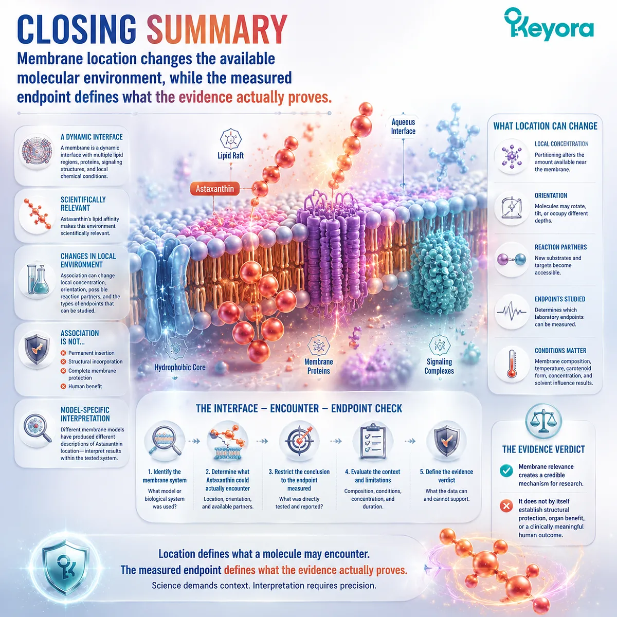
Closing Summary
Membrane location changes the available molecular environment, while the measured endpoint defines what the evidence actually proves
A membrane is not merely a lipid wall or another address inside the body. It is a dynamic interface containing different lipid regions, membrane proteins, signaling structures, and local chemical conditions.
Astaxanthin’s lipid affinity makes this environment scientifically relevant. Membrane association can change its local concentration, orientation, possible reaction partners, and the kinds of laboratory endpoints researchers can investigate.
However, association is not the same as permanent insertion, structural incorporation, complete membrane protection, or human benefit. Different membrane models have produced different descriptions of Astaxanthin location, reinforcing the need for model-specific interpretation.
The Interface – Encounter – Endpoint Check provides the practical verdict. Identify the membrane system, determine what Astaxanthin could actually encounter, and restrict the conclusion to the endpoint measured.
Astaxanthin’s membrane relevance creates a credible mechanism for research. It does not by itself establish structural protection, organ benefit, or a clinically meaningful human outcome.
This article is for educational and informational purposes only. It does not provide medical advice, diagnosis, treatment, cure, prevention, disease outcome claims, hormone restoration claims, fertility outcome claims, or formula-specific clinical efficacy claims.