
How Does Astaxanthin Interact With Cell Membranes?
Keyora Research Q&A Library
This is part of the Keyora Research Q&A Series, derived from Keyora Astaxanthin Research Journal Series.
Within the Keyora Astaxanthin Researcn framework, this Q&A translates complex astaxanthin biology into reader-friendly, evidence-bound answers, focusing on natural astaxanthin identity, molecular structure, antioxidant and redox mechanisms, membrane lipid interaction, mitochondrial resilience, inflammatory signaling pathways, human evidence interpretation, and the scientific principles behind responsible supplementation.
First published by Keyora Research Journal: www.keyorahealth.com
Direct Answer
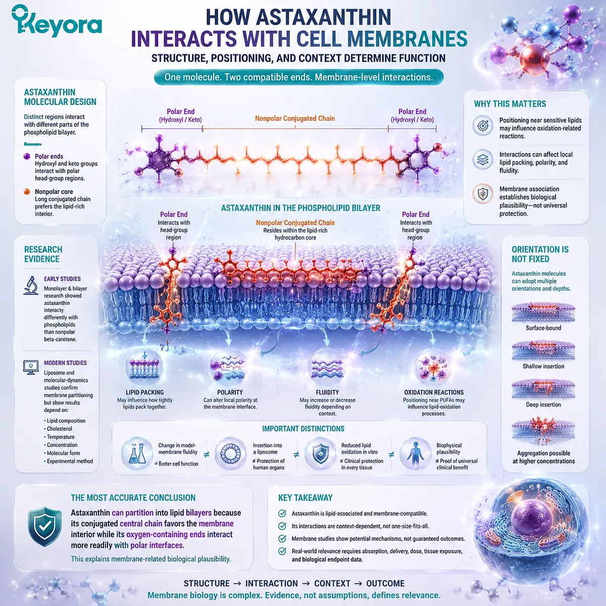
Astaxanthin can interact with cell membranes because different regions of its molecular structure are compatible with different parts of a phospholipid bilayer. Its long conjugated central chain favors the membrane’s lipid-rich interior, while the hydroxyl- and keto-containing terminal rings can interact more readily with the relatively polar regions near phospholipid head groups.
This does not mean that every astaxanthin molecule assumes one permanent position. Membrane studies support astaxanthin partitioning into phospholipid systems, but its depth, angle, movement, aggregation state, and effects on bilayer properties can change with membrane composition, cholesterol, temperature, concentration, molecular form, and the experimental method. Early monolayer and bilayer research found that astaxanthin interacted differently with phospholipids than nonpolar beta-carotene. More recent liposome and molecular-dynamics studies also show that membrane behavior is context-dependent rather than fixed.
Membrane association may place astaxanthin near oxidation-sensitive lipids and influence local lipid packing, polarity, fluidity, or oxidation-related reactions. However, these outcomes are not interchangeable. A change in model-membrane fluidity does not automatically prove better cell function, and insertion into a liposome does not prove protection of human organs.
The most accurate conclusion is:
Astaxanthin can partition into lipid bilayers because its conjugated central chain favors the membrane interior while its oxygen-containing ends interact more readily with polar interfaces.
This establishes membrane-related biological plausibility. It does not prove one universal orientation, complete membrane coverage, or clinical protection of every human tissue.
Why Astaxanthin Can Associate With Lipid Bilayers
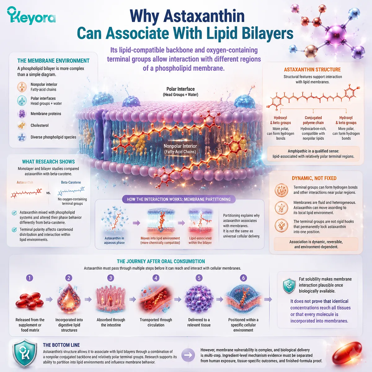
Its lipid-compatible backbone and oxygen-containing terminal groups allow interaction with different regions of a phospholipid membrane
A phospholipid bilayer contains a relatively nonpolar interior formed by fatty-acid chains and more polar interfaces formed by phospholipid head groups and surrounding water. Membrane proteins, cholesterol, and different phospholipid species make the complete environment considerably more complex than a simple diagram.
Astaxanthin contains a long, hydrocarbon-rich conjugated chain. This central region is compatible with nonpolar lipid environments and can interact with phospholipid fatty-acid chains. The terminal rings contain hydroxyl and keto groups, creating regions that can participate more readily in polar interactions.
This structural combination is sometimes described as amphipathic. The term requires qualification because astaxanthin remains strongly fat-soluble and does not behave like a freely water-soluble molecule. A more precise description is that astaxanthin is lipid-associated but contains relatively polar terminal regions.
Primary monolayer and bilayer research compared astaxanthin with beta-carotene, which lacks oxygen-containing terminal groups. Astaxanthin mixed with phospholipid systems and altered their phase behavior differently from beta-carotene, supporting the conclusion that terminal polarity affects carotenoid distribution and interaction within lipid environments.
The interaction can be understood as membrane partitioning. Astaxanthin moves into a lipid environment because that environment is more chemically compatible with most of the molecule than the surrounding aqueous phase.
Partitioning is not the same as universal cellular delivery. After oral consumption, astaxanthin must still be:
-
released from the supplement or food matrix
-
incorporated into digestive lipid structures
-
absorbed through the intestine
-
transported through circulation
-
delivered to a relevant tissue
-
positioned within a specific cellular environment
Fat solubility explains why membrane interaction is plausible after astaxanthin becomes biologically available. It does not prove that identical concentrations reach all tissues or that every circulating molecule becomes incorporated into a cell membrane.
The terminal groups also do not function as rigid hooks that permanently lock astaxanthin into one position. Hydrogen bonding and other molecular interactions may contribute to association near polar membrane regions, but membranes are dynamic, and astaxanthin can move in response to its local lipid environment.
What Membrane Models Suggest About Astaxanthin’s Behavior
Biophysical studies support membrane insertion and several possible behaviors rather than one fixed arrangement
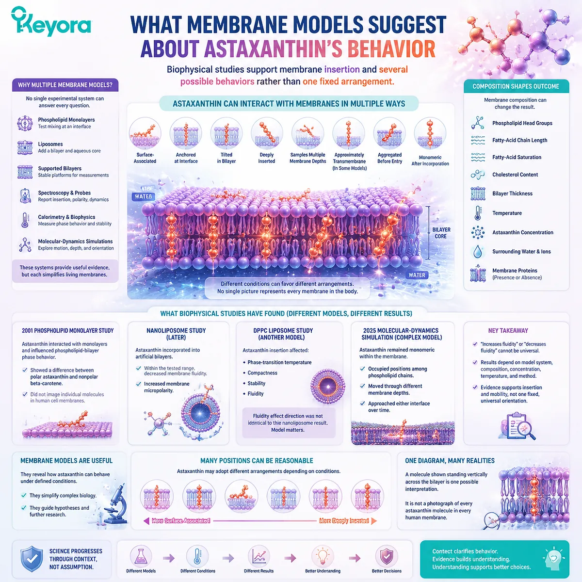
Researchers use several types of membrane models because no single experimental system can answer every question.
A phospholipid monolayer can show whether a carotenoid mixes with lipids at an interface. A liposome adds a complete lipid bilayer and an enclosed aqueous space. Supported bilayers, spectroscopy, fluorescence probes, calorimetry, and molecular-dynamics simulations can examine additional properties such as insertion, molecular movement, phase transitions, polarity, and possible orientation.
These systems provide useful evidence, but each simplifies living membranes.
The 2001 phospholipid study found that astaxanthin interacted with monolayers and influenced phospholipid-bilayer phase behavior. The study supported a difference between polar astaxanthin and nonpolar beta-carotene, but it did not photograph individual astaxanthin molecules inside human cell membranes.
A later nanoliposome study incorporated astaxanthin into artificial bilayers and used fluorescent probes to examine membrane properties. Within the tested concentration range, astaxanthin incorporation decreased measured membrane fluidity and increased membrane micropolarity. Those results apply to that liposome composition, concentration range, and measurement method.
Another DPPC liposome study reported that astaxanthin insertion affected phase-transition temperature, compactness, stability, and fluidity. The direction of the reported fluidity effect was not identical to the earlier nanoliposome result. This difference is scientifically useful because it demonstrates that “astaxanthin increases membrane fluidity” or “astaxanthin decreases membrane fluidity” cannot be treated as universal statements.
Membrane composition can change the outcome through differences in:
-
phospholipid head groups
-
fatty-acid chain length
-
fatty-acid saturation
-
cholesterol content
-
bilayer thickness
-
temperature
-
astaxanthin concentration
-
surrounding water and ions
-
the presence or absence of membrane proteins
A 2025 molecular-dynamics investigation used a more compositionally complex membrane model. In that simulation, astaxanthin remained monomeric within the membrane, occupied positions among phospholipid hydrocarbon chains, and moved through different membrane depths. The modeled molecules could approach either membrane interface over time rather than remaining in one motionless location.
This newer model is important because it challenges overly rigid diagrams. It supports membrane insertion and substantial molecular mobility in that simulated environment, but it remains a computational model with defined lipids, force fields, temperature, and simulation time.
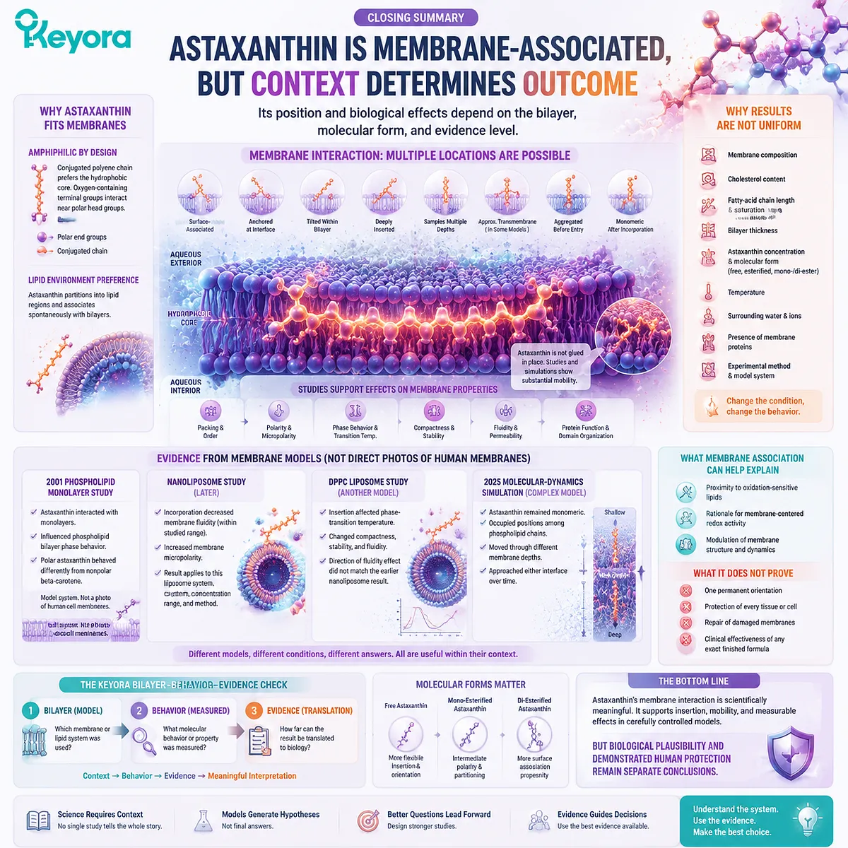
Several descriptions may therefore be reasonable under different conditions:
-
surface-associated
-
anchored near an interface
-
tilted within the bilayer
-
deeply inserted
-
able to sample multiple membrane depths
-
approximately transmembrane in a particular model
-
aggregated before membrane entry
-
monomeric after incorporation
A diagram showing one molecule standing vertically across the bilayer represents one possible structural interpretation. It should not be presented as a photograph of every astaxanthin molecule in every human membrane.
Why Membrane Association May Matter for Redox Biology
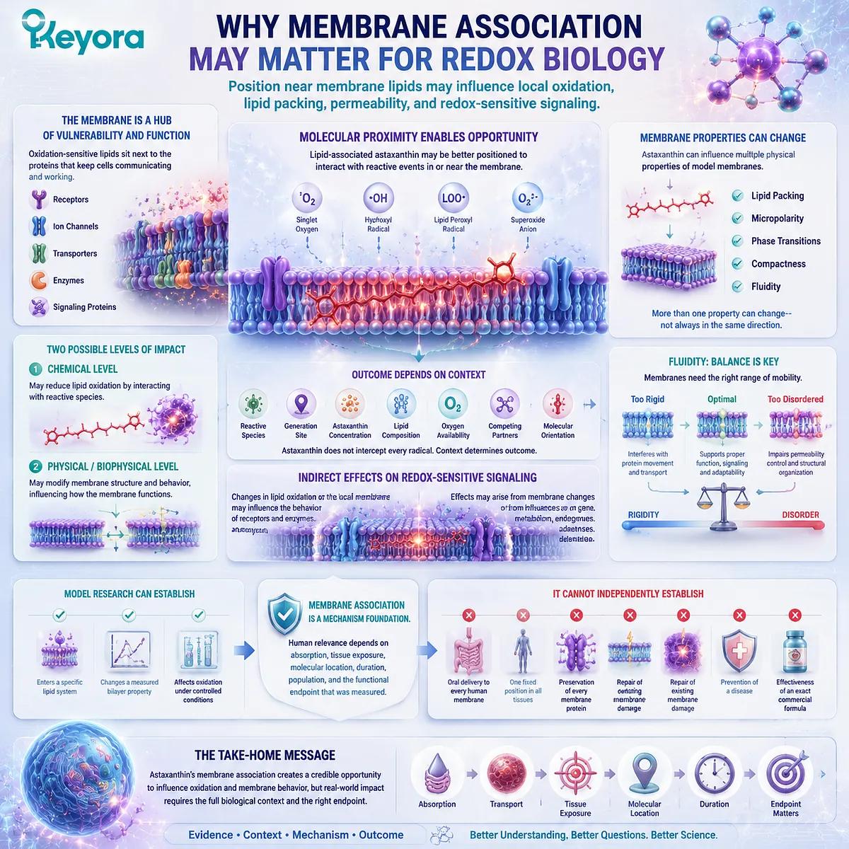
Position near membrane lipids may influence local oxidation, lipid packing, permeability, and redox-sensitive signaling
Cell membranes contain oxidation-sensitive lipids positioned beside receptors, channels, transporters, and signaling proteins. Astaxanthin’s ability to associate with lipid bilayers creates a credible reason to study it near these targets.
The first possible benefit is molecular proximity. A lipid-associated carotenoid may be better positioned to encounter selected reactive events occurring within or near a membrane than a molecule restricted mainly to an aqueous environment.
This does not mean that astaxanthin intercepts every radical. The outcome depends on:
-
which reactive species is present
-
where it is generated
-
the astaxanthin concentration
-
the surrounding lipid composition
-
oxygen availability
-
competing reaction partners
-
the molecular orientation at that moment
The second possible effect involves the physical membrane environment. Incorporating astaxanthin into model bilayers has altered lipid packing, micropolarity, phase transitions, compactness, and fluidity. These changes show that astaxanthin can influence more than a single chemical oxidation reaction. They also show why “membrane stabilization” requires a specific definition.
Stabilization could refer to:
-
reduced leakage
-
preserved bilayer organization
-
resistance to temperature-related transition
-
lower lipid oxidation
-
altered fluidity
-
greater structural compactness
These endpoints cannot be assumed to move in the same direction or to be beneficial under every condition.
Membrane fluidity provides an important example. Cell membranes require an appropriate range of mobility. Excessive rigidity can interfere with protein movement and transport, while excessive disorder can impair permeability control and structural organization. The goal is not maximum fluidity or minimum fluidity, but context-appropriate membrane behavior.
Membrane association may also affect redox-sensitive signaling indirectly. Changes in lipid oxidation or local bilayer organization may alter the environment surrounding receptors and enzymes. However, a pathway change in cultured cells cannot be attributed automatically to direct membrane insertion because astaxanthin may also influence gene expression, metabolism, or endogenous defense systems.
Model research can establish:
-
that astaxanthin enters a particular lipid system
-
that it changes a measured bilayer property
-
that it affects oxidation under controlled conditions
It cannot independently establish:
-
oral delivery to every human membrane
-
one fixed position in all tissues
-
preservation of every membrane protein
-
repair of existing membrane damage
-
prevention of a disease
-
effectiveness of an exact commercial formula
Membrane association is therefore a mechanism foundation. Human relevance still depends on absorption, tissue exposure, molecular location, duration, population, and the functional endpoint that was measured.
Use the Keyora Bilayer – Behavior – Evidence Check
Three questions separate a model of membrane interaction from a claim of whole-body protection
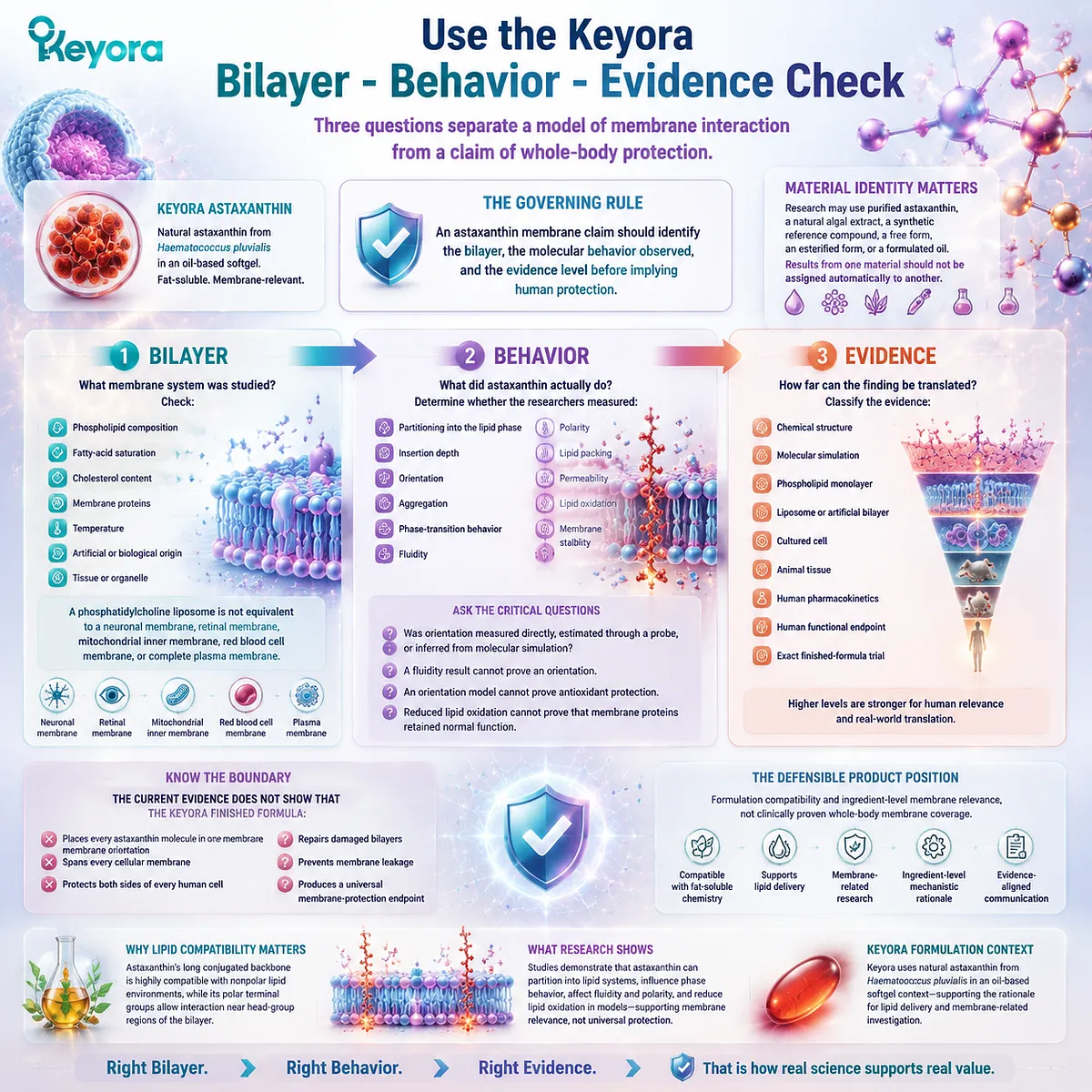
The Keyora Bilayer – Behavior – Evidence Check provides a practical method for evaluating membrane claims about astaxanthin.
1. Bilayer
What membrane system was studied?
Check:
-
phospholipid composition
-
fatty-acid saturation
-
cholesterol content
-
membrane proteins
-
temperature
-
artificial or biological origin
-
tissue or organelle
A phosphatidylcholine liposome is not equivalent to a neuronal membrane, retinal membrane, mitochondrial inner membrane, red blood cell membrane, or complete plasma membrane.
2. Behavior
What did astaxanthin actually do?
Determine whether the researchers measured:
-
partitioning into the lipid phase
-
insertion depth
-
orientation
-
aggregation
-
phase-transition behavior
-
fluidity
-
polarity
-
lipid packing
-
permeability
-
lipid oxidation
-
membrane stability
Also ask whether orientation was measured directly, estimated through a probe, or inferred from molecular simulation.
A fluidity result cannot prove an orientation. An orientation model cannot prove antioxidant protection. Reduced lipid oxidation cannot prove that membrane proteins retained normal function.
3. Evidence
How far can the finding be translated?
Classify the evidence:
-
chemical structure
-
molecular simulation
-
phospholipid monolayer
-
liposome or artificial bilayer
-
cultured cell
-
animal tissue
-
human pharmacokinetics
-
human functional endpoint
-
exact finished-formula trial
The governing rule is:
An astaxanthin membrane claim should identify the bilayer, the molecular behavior observed, and the evidence level before implying human protection.
Material identity also matters. Research may use purified astaxanthin, a natural algal extract, a synthetic reference compound, a free form, an esterified form, or a formulated oil. Results from one material should not be assigned automatically to another.
Keyora uses natural astaxanthin from Haematococcus pluvialis in an oil-based softgel context. This is compatible with astaxanthin’s fat-soluble chemistry and supports the rationale for lipid delivery and membrane-related research.
The supplied Keyora knowledge system also establishes an important boundary: the formula has extensive ingredient-level rationale, but an exact finished-formula human clinical trial was not established in the supplied corpus.
The current evidence therefore does not show that the Keyora finished formula:
-
places every astaxanthin molecule in one membrane orientation
-
spans every cellular membrane
-
protects both sides of every human cell
-
repairs damaged bilayers
-
prevents membrane leakage
-
produces a universal membrane-protection endpoint
The defensible product position is formulation compatibility and ingredient-level membrane relevance, not clinically proven whole-body membrane coverage.
Closing Summary
Astaxanthin is membrane-associated, but its position and biological effects depend on the bilayer, molecular form, and evidence level
Astaxanthin can associate with phospholipid bilayers because its conjugated central chain favors lipid environments while its oxygen-containing terminal groups interact more readily near polar interfaces.
Membrane studies support insertion, movement through different bilayer depths, and effects on properties such as packing, polarity, phase behavior, compactness, and fluidity. The results are not uniform because membrane composition, cholesterol, concentration, temperature, molecular form, and experimental method change the observed behavior.
Membrane association may position astaxanthin near oxidation-sensitive lipids and provide a rationale for membrane-centered redox research. It does not prove one permanent orientation, protection of every tissue, repair of damaged membranes, or clinical effectiveness of an exact finished formula.
Use the Keyora Bilayer – Behavior – Evidence Check. Identify the membrane model, determine what molecular behavior was measured, and confirm how far the evidence can be translated.
Astaxanthin’s membrane interaction is scientifically meaningful, but biological plausibility and demonstrated human protection remain separate conclusions.
This article is for educational and informational purposes only. It does not provide medical advice, diagnosis, treatment, cure, prevention, disease outcome claims, hormone restoration claims, fertility outcome claims, or formula-specific clinical efficacy claims.