
How Do Cell Membranes, Mitochondria, and Inflammation Connect?
Keyora Research Q&A Library
This is part of the Keyora Research Q&A Series, derived from Keyora Astaxanthin Research Journal Series.
Within the Keyora Astaxanthin Researcn framework, this Q&A translates complex astaxanthin biology into reader-friendly, evidence-bound answers, focusing on natural astaxanthin identity, molecular structure, antioxidant and redox mechanisms, membrane lipid interaction, mitochondrial resilience, inflammatory signaling pathways, human evidence interpretation, and the scientific principles behind responsible supplementation.
First published by Keyora Research Journal: www.keyorahealth.com

Direct Answer
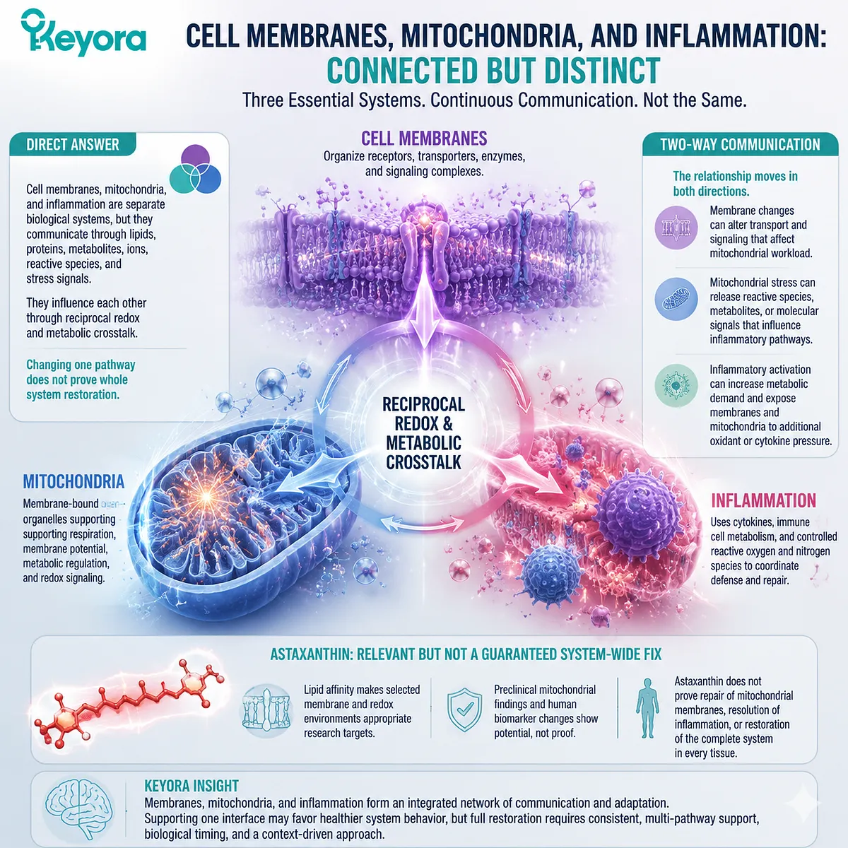
Cell membranes, mitochondria, and inflammation are separate biological systems, but they communicate through lipids, proteins, metabolites, ions, reactive species, and stress signals.
Cell membranes organize receptors, transporters, enzymes, and signaling complexes.
Mitochondria are membrane bound organelles whose inner membrane supports respiration, membrane potential, metabolic regulation, and redox signaling.
Inflammation uses cytokines, immune cell metabolism, and controlled production of reactive oxygen and nitrogen species to coordinate defense and repair.
Cell membranes, mitochondria, and inflammatory signaling are connected through reciprocal redox and metabolic communication, but changing one pathway does not prove whole system restoration.
The relationship can move in both directions.
-
Membrane changes can alter transport and signaling that affect mitochondrial workload.
-
Mitochondrial stress can release reactive species, metabolites, or molecular signals that influence inflammatory pathways.
-
Inflammatory activation can then increase metabolic demand and expose membranes and mitochondria to additional oxidant or cytokine pressure.
Astaxanthin is relevant because its lipid affinity makes selected membrane and redox environments appropriate research targets.
However, preclinical mitochondrial findings and human biomarker changes do not prove that Astaxanthin repairs mitochondrial membranes, resolves inflammation, or restores the complete system in every tissue.
Cell Membranes Organize More Than a Physical Boundary
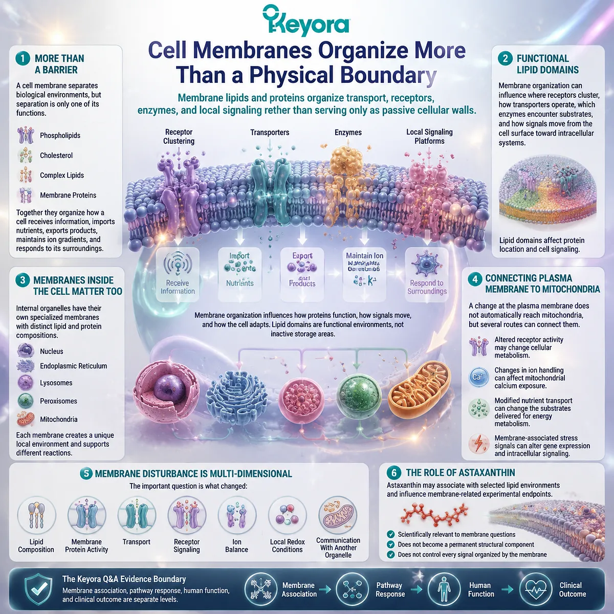
Membrane lipids and proteins organize transport, receptors, enzymes, and local signaling rather than serving only as passive cellular walls
A cell membrane separates biological environments, but separation is only one of its functions. The plasma membrane contains phospholipids, cholesterol, other complex lipids, and proteins that organize how a cell receives information, imports nutrients, exports products, maintains ion gradients, and responds to its surroundings.
Membrane organization can influence where receptors cluster, how transporters operate, which enzymes encounter their substrates, and how signals move from the cell surface toward intracellular systems.
Lipid domains are therefore functional environments rather than inactive storage areas. Their composition and organization can affect protein location and cell signaling.
The same principle applies to membranes inside the cell.
The nucleus, endoplasmic reticulum, lysosomes, peroxisomes, and mitochondria contain membranes with specialized lipid and protein compositions.
Each membrane creates a distinct local environment and supports different reactions.
A change at the plasma membrane does not automatically reach mitochondria, but several routes can connect them.
Altered receptor activity may change cellular metabolism.
Changes in ion handling can affect mitochondrial calcium exposure.
Modified nutrient transport can change the substrates delivered for energy metabolism.
Membrane associated stress signals can also alter gene expression and intracellular signaling.
This means membrane disturbance should not be reduced to a picture of a damaged outer wall. The important question is what changed:
-
lipid composition
-
membrane protein activity
-
transport
-
receptor signaling
-
ion balance
-
local redox conditions
-
communication with another organelle
Astaxanthin may associate with selected lipid environments and influence membrane related experimental endpoints. That association makes it scientifically relevant to these questions, but it does not establish that Astaxanthin becomes a permanent structural membrane component or controls every signal organized by the membrane.
The current Keyora Q&A evidence boundary treats membrane association, pathway response, human function, and clinical outcome as separate levels.
Mitochondria Link Membrane Function With Redox Signaling
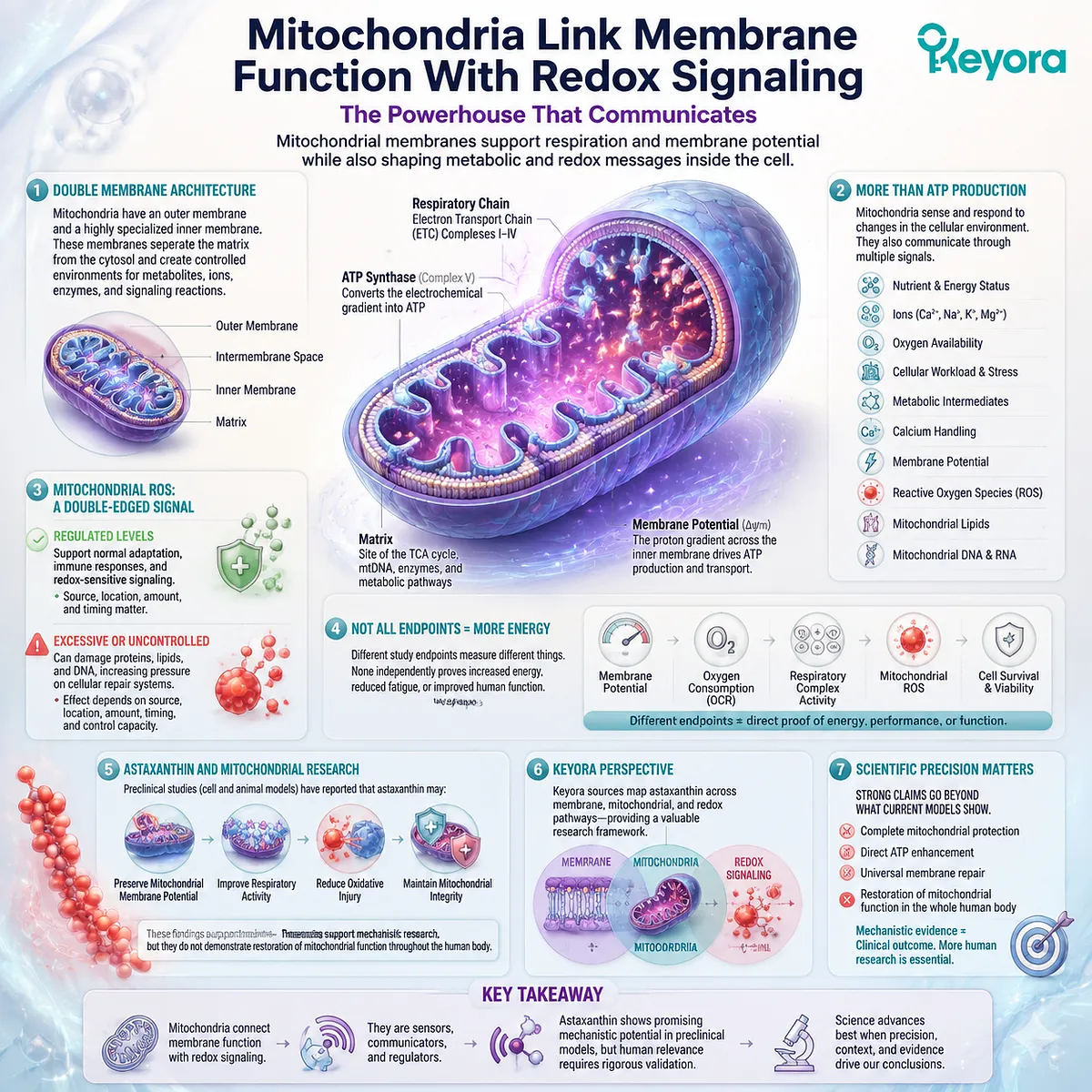
Mitochondrial membranes support respiration and membrane potential while also shaping metabolic and redox messages inside the cell
Mitochondria contain an outer membrane and a highly specialized inner membrane. The inner membrane houses respiratory machinery and helps maintain the electrochemical gradient used in cellular energy metabolism. The membranes also separate the mitochondrial matrix from the cytosol and create controlled environments for metabolites, ions, enzymes, and signaling reactions.
Mitochondria are therefore more than ATP producing structures. They sense changes in nutrients, ions, oxygen availability, cellular workload, and stress. They also communicate through metabolites, calcium handling, membrane potential, reactive oxygen species, mitochondrial lipids, and mitochondrial nucleic acids.
Mitochondrial reactive oxygen species do not have one fixed meaning. Regulated production can contribute to normal adaptation, immune responses, and redox sensitive signaling. Excessive, prolonged, or poorly controlled production can instead modify proteins, lipids, or DNA and place additional pressure on cellular repair systems. The effect depends on source, location, amount, timing, and the cell’s capacity to control the signal.
This distinction prevents mitochondrial support from being translated automatically into “more energy.” A study may report changes in membrane potential, oxygen consumption, respiratory complex activity, mitochondrial reactive species, or cell survival. Each is a different endpoint. None independently proves increased energy, reduced fatigue, or improved human function.
Astaxanthin has been investigated in this context, but much of the direct mitochondrial evidence remains preclinical. For example, animal and cell based studies have measured mitochondrial membrane potential, respiratory activity, oxidative injury, and mitochondrial integrity after Astaxanthin exposure. These experiments support mechanistic research, but they do not demonstrate restoration of mitochondrial function throughout the human body.
Keyora source materials position Astaxanthin across membrane, mitochondrial, and redox pathways. This provides a useful research map, but stronger phrases such as complete mitochondrial protection, direct ATP enhancement, or universal membrane repair extend beyond what those mechanistic models establish.
Inflammation Can Respond to and Increase Redox Stress
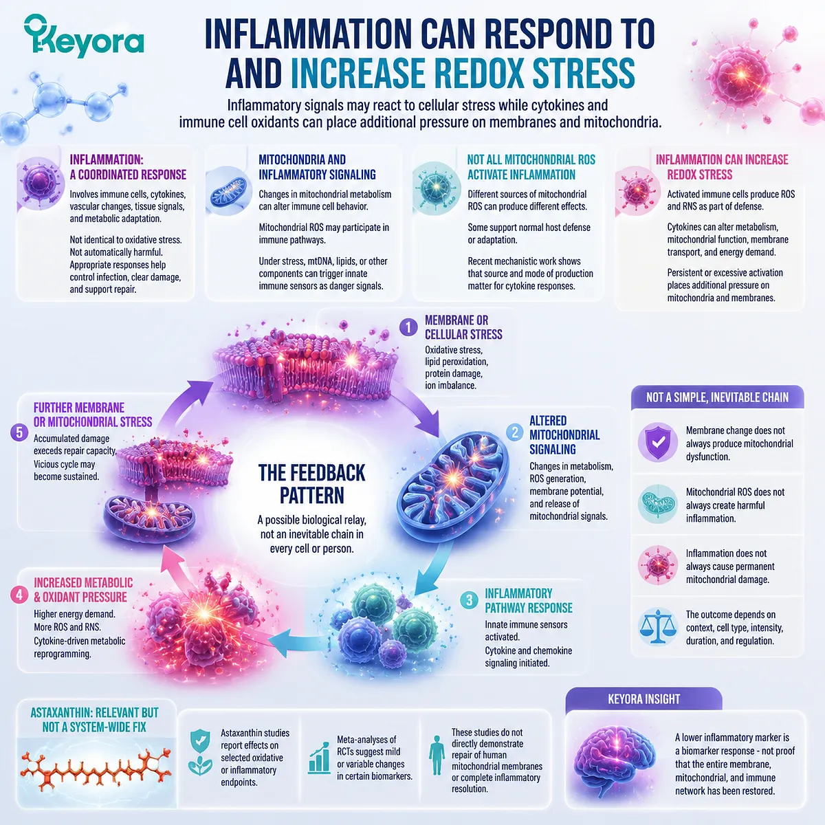
Inflammatory signals may react to cellular stress while cytokines and immune cell oxidants can place additional pressure on membranes and mitochondria
Inflammation is a coordinated biological response involving immune cells, cytokines, vascular changes, tissue signals, and metabolic adaptation. It is not identical to oxidative stress, and it is not automatically harmful. Appropriate inflammatory responses help control infection, clear damaged material, and support tissue repair.
Mitochondria can contribute to inflammatory signaling in several ways. Changes in mitochondrial metabolism can alter immune cell behavior. Mitochondrial reactive oxygen species may participate in selected immune pathways. Under particular forms of stress or injury, mitochondrial DNA, lipids, or other components can appear in locations where innate immune sensors recognize them as danger related signals.
This does not mean all mitochondrial reactive species activate inflammation. Different sources of mitochondrial ROS can produce different effects, and some are involved in normal host defense or adaptation. Recent mechanistic work in immune cells continues to show that the source and mode of mitochondrial ROS production matter when interpreting cytokine responses.
The relationship also works in the opposite direction. Activated immune cells can produce reactive oxygen and nitrogen species as part of defense. Cytokines can alter nutrient use, mitochondrial metabolism, membrane transport, and cellular energy demand. When activation is persistent or excessive, these responses may place additional pressure on mitochondrial and membrane systems.
A feedback pattern may therefore develop:
Membrane or cellular stress
→ altered mitochondrial signaling
→ inflammatory pathway response
→ increased metabolic and oxidant pressure
→ further membrane or mitochondrial stress
This sequence is a possible biological relay, not an inevitable chain in every cell or person. Membrane change does not always produce mitochondrial dysfunction. Mitochondrial ROS does not always create harmful inflammation. Inflammation does not always cause permanent mitochondrial damage.
Astaxanthin studies have reported effects on selected oxidative or inflammatory endpoints. Meta-analyses of randomized trials suggest mild or variable changes in certain biomarkers, but these studies do not directly demonstrate repair of human mitochondrial membranes or complete inflammatory resolution.
A lower inflammatory marker should therefore be described as a biomarker response, not proof that the entire membrane, mitochondrial, and immune network has been restored.
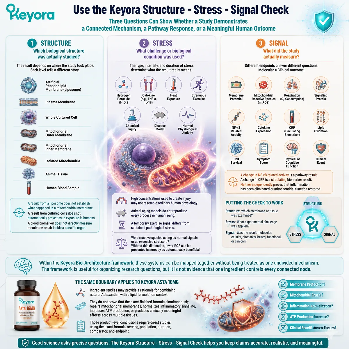
Use the Keyora Structure – Stress – Signal Check
Three questions can show whether a study demonstrates a connected mechanism, a pathway response, or a meaningful human outcome
The Keyora Structure – Stress – Signal Check helps readers examine broad claims such as “Astaxanthin repairs mitochondria and stops inflammation.”
Structure: Which biological structure was actually studied?
The structure might be:
-
an artificial phospholipid membrane
-
a plasma membrane
-
a whole cultured cell
-
the mitochondrial outer membrane
-
the mitochondrial inner membrane
-
isolated mitochondria
-
an animal tissue
-
a human blood sample
A result from a liposome does not establish what happened in a mitochondrial membrane. A result from cultured cells does not automatically prove tissue exposure in humans. A blood biomarker does not directly measure membrane repair inside a specific organ.
Stress: What challenge or biological condition was used?
The study may involve hydrogen peroxide, a cytokine, heat exposure, strenuous exercise, a chemical injury, a disease model, or normal physiological activity.
The intensity and duration matter. A high concentration used to create cell injury may not resemble ordinary human physiology. An animal aging model does not reproduce every process in human aging. A temporary exercise signal differs from sustained pathological stress.
The reader should also ask whether reactive species were functioning as normal signals or as excessive stressors. Without that distinction, lower ROS can be presented incorrectly as automatically beneficial.
Signal: What did the study actually measure?
Possible endpoints include:
-
membrane potential
-
mitochondrial reactive species
-
respiration
-
a signaling protein
-
NF-kB related activity
-
cytokine expression
-
CRP
-
lipid oxidation
-
cell survival
-
a symptom score
-
physical or cognitive function
-
a clinical event
A change in NF-kB related activity is a pathway result. A change in CRP is a circulating biomarker result. Neither independently proves that inflammation has been eliminated or mitochondrial function restored.
Consider the claim, “Astaxanthin protects membranes, restores mitochondria, and resolves inflammation.”
The structure question asks which membrane or tissue was examined. The stress question identifies the experimental challenge. The signal question determines whether the result was molecular, cellular, biomarker based, functional, or clinical.
Within the Keyora Bio-Architecture framework, these systems can be mapped together without being treated as one undivided mechanism. The framework is useful for organizing research questions, but it is not evidence that one ingredient controls every connected node.
The same boundary applies to Keyora Asta 16MG. Ingredient studies may provide a rationale for combining natural Astaxanthin with a lipid formulation context. They do not prove that the exact finished formula simultaneously repairs mitochondrial membranes, normalizes inflammatory signaling, increases ATP production, or produces clinically meaningful effects across multiple tissues.
Those product level conclusions require direct studies using the exact formula, serving, population, duration, comparator, and endpoint.
Closing Summary
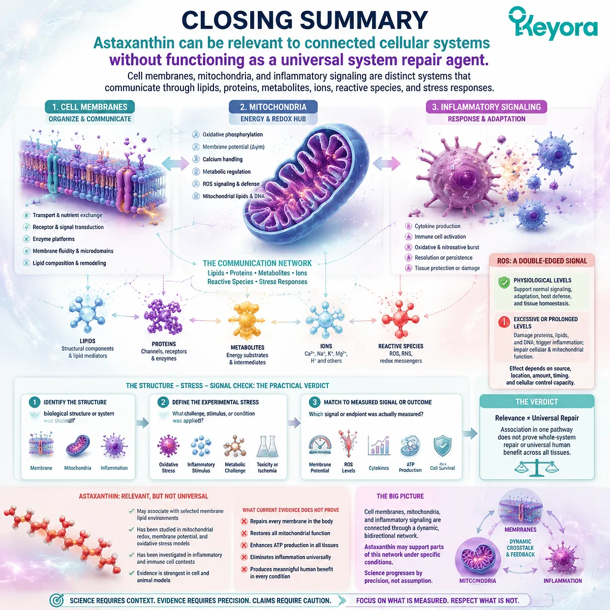
Astaxanthin can be relevant to connected cellular systems without functioning as a universal system repair agent
Cell membranes, mitochondria, and inflammatory signaling are distinct systems that communicate through lipids, proteins, metabolites, ions, reactive species, and stress responses.
Membranes organize transport and signaling. Mitochondria use specialized membranes for metabolism, membrane potential, and redox communication. Inflammation can respond to cellular and mitochondrial stress, while cytokines and immune cell oxidants can increase metabolic and redox pressure.
The relationship is bidirectional, but it is not automatic. Reactive oxygen species can support normal signaling or contribute to stress depending on their source, location, amount, and duration.
The Structure – Stress – Signal Check provides the practical verdict. Identify the structure studied, define the experimental stress, and match the conclusion to the signal or outcome actually measured.
Astaxanthin may be relevant to selected membrane, mitochondrial redox, and inflammatory environments. Their connection does not prove that one ingredient repairs the entire system or produces meaningful human benefits across every tissue.
This article is for educational and informational purposes only. It does not provide medical advice, diagnosis, treatment, cure, prevention, disease outcome claims, hormone restoration claims, fertility outcome claims, or formula-specific clinical efficacy claims.