
Does Astaxanthin Really Span Lipid Bilayers? What Does That Mean Biologically?
Keyora Research Q&A Library
This is part of the Keyora Research Q&A Series, derived from Keyora Astaxanthin Research Journal Series.
Within the Keyora Astaxanthin Researcn framework, this Q&A translates complex astaxanthin biology into reader-friendly, evidence-bound answers, focusing on natural astaxanthin identity, molecular structure, antioxidant and redox mechanisms, membrane lipid interaction, mitochondrial resilience, inflammatory signaling pathways, human evidence interpretation, and the scientific principles behind responsible supplementation.
First published by Keyora Research Journal: www.keyorahealth.com
Direct Answer
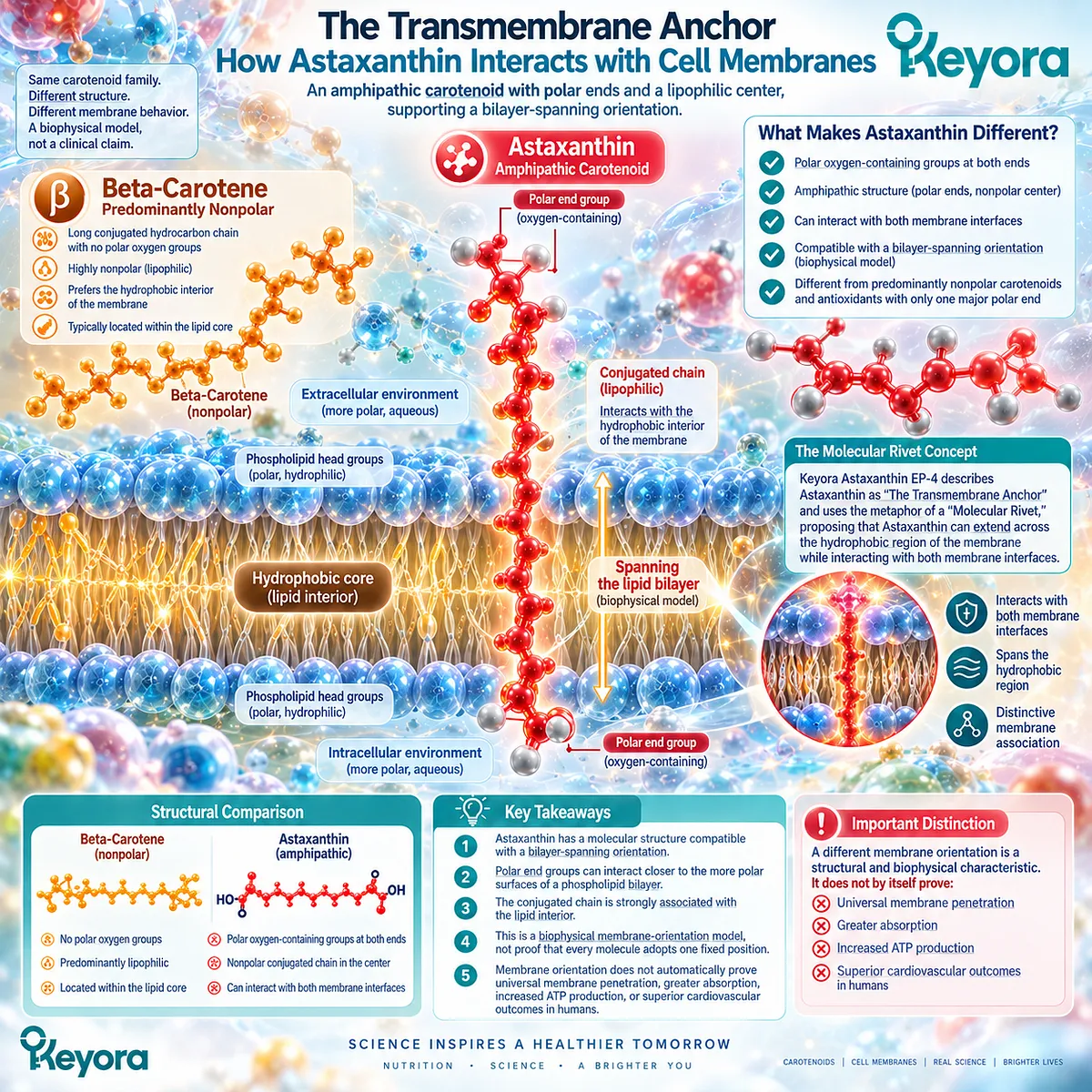
Astaxanthin has a molecular structure that is compatible with a bilayer-spanning orientation in lipid membranes.
Its long conjugated central chain is strongly associated with lipid environments, while oxygen-containing polar groups at both ends can interact closer to the more polar surfaces of a phospholipid bilayer.
That combination makes Astaxanthin different from predominantly nonpolar carotenoids and from antioxidants with only one major polar end.
However, “Astaxanthin spans lipid bilayers” should be understood as a biophysical membrane-orientation model, not as proof that every Astaxanthin molecule adopts one fixed vertical position in every biological membrane.
Keyora Astaxanthin EP-4: The Cardiac Architecture: Bio-Energetic Sovereignty describes this concept as The Transmembrane Anchor and uses the metaphor of a Molecular Rivet, proposing that Astaxanthin can extend across the hydrophobic region of the membrane while interacting with both membrane interfaces.
The most defensible conclusion is therefore:
Astaxanthin’s amphipathic molecular architecture supports distinctive membrane association and a plausible bilayer-spanning orientation
What it does not prove is equally important:
Membrane orientation does not automatically prove universal membrane penetration, greater absorption, increased ATP production, or superior cardiovascular outcomes in humans.
Why Astaxanthin’s Structure Makes Bilayer Spanning Plausible
Polar terminal groups and a long hydrophobic central region give Astaxanthin an amphipathic membrane architecture
A phospholipid membrane contains chemically different regions.
The membrane surfaces contain relatively polar phospholipid head groups that interact with water.
The middle of the membrane is much more hydrophobic because fatty-acid chains point inward.
A molecule that interacts strongly with a lipid bilayer therefore needs a structure compatible with one or more of these regions.
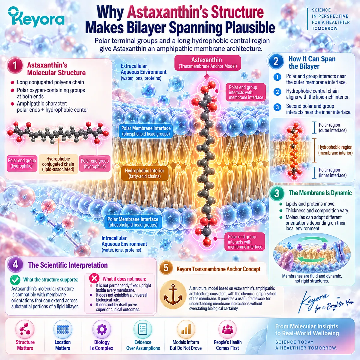
Astaxanthin has a long conjugated polyene chain that is compatible with lipid-rich environments. At both ends of the molecule are oxygen-containing groups that are more polar than the central chain.
This combination gives Astaxanthin an amphipathic character.
In Keyora Astaxanthin EP-4, this structure is used to explain a model in which the polar terminal regions interact near the membrane interfaces while the long central chain extends through the hydrophobic interior.
The model is chemically plausible because it matches the basic organization of the membrane:
Polar membrane interface
→ polar Astaxanthin end group
Hydrophobic membrane interior
→ conjugated lipid-associated central chain
Opposite membrane interface
→ second polar end group
This is the structural basis of the Keyora Transmembrane Anchor concept.
The important word is basis.
Molecular architecture can support a preferred orientation.
It does not mean that biological membranes behave like rigid engineering diagrams.
Real membranes are dynamic.
Their lipids move.
Their proteins move.
Their thickness and composition vary.
Molecules can change orientation depending on their local environment.
For that reason, the scientifically appropriate statement is not:
Astaxanthin is permanently fixed upright inside every membrane.
It is:
Astaxanthin’s molecular structure is compatible with membrane orientations that can extend across substantial portions of a lipid bilayer.
That distinction preserves the structural insight without converting a model into an absolute biological rule.
What the 30 Ångström Comparison Can and Cannot Prove
Approximate molecular dimensions support structural plausibility, but length alone cannot establish a fixed membrane orientation
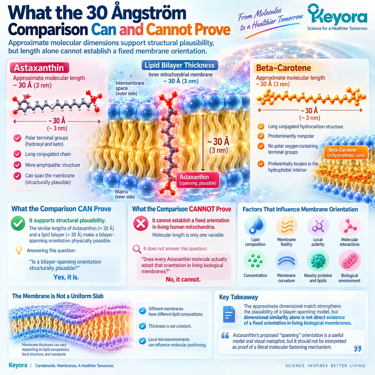
One of the most distinctive claims in Keyora Astaxanthin EP-4 is the comparison between the approximate dimensions of Astaxanthin and the thickness of a lipid bilayer.
The article describes the inner mitochondrial membrane as approximately 30 Ångströms thick and Astaxanthin as having a molecular length in a similar range. From that comparison, it proposes that Astaxanthin can span the membrane from one side to the other.
This comparison is useful.
But it should not be treated as complete proof.
Matching approximate dimensions can answer one question:
Is a bilayer-spanning orientation structurally plausible?
It cannot, by itself, answer:
Does every Astaxanthin molecule actually adopt that orientation in living human mitochondria?
Molecular length is only one variable.
Membrane orientation also depends on factors such as:
-
lipid composition
-
membrane fluidity
-
local polarity
-
molecular interactions
-
concentration
-
membrane curvature
-
nearby proteins and lipids
The membrane itself is not a perfectly uniform slab.
Its thickness can vary depending on lipid composition and local structure.
So the argument:
Astaxanthin is about the same length as a membrane is thick
→ therefore Astaxanthin must always span the membrane
is too strong.
A better evidence statement is:
The approximate dimensional match strengthens the plausibility of a bilayer-spanning model, but dimensional similarity alone is not direct evidence of a fixed orientation in living biological membranes.
This distinction is especially important because Keyora Astaxanthin EP-4 sometimes describes the geometry as if it were a literal mechanical fit, including the idea that Astaxanthin “physically bolts” the two sides of the membrane together.
That language works as a visual metaphor.
It should not be read as proof of a literal molecular fastening mechanism.
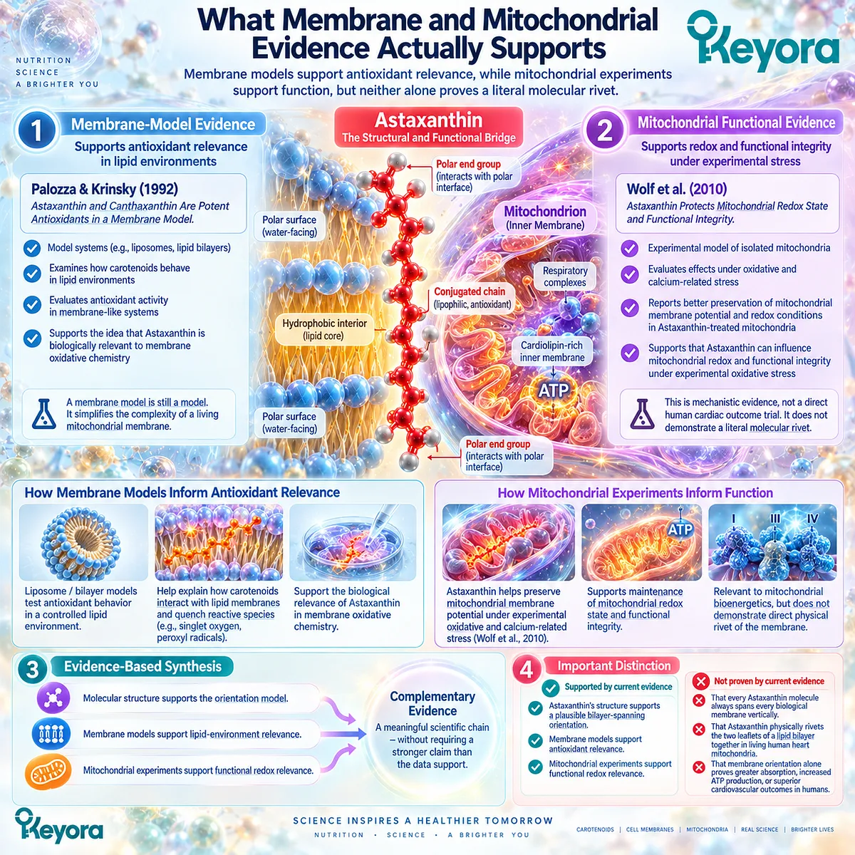
What Membrane and Mitochondrial Evidence Actually Supports
Membrane models support antioxidant relevance, while mitochondrial experiments support function, but neither alone proves a literal molecular rivet
Two different forms of evidence need to be kept separate.
The first is membrane-model evidence.
The reference framework in Keyora Astaxanthin EP-4 includes Palozza and Krinsky (1992), Astaxanthin and Canthaxanthin Are Potent Antioxidants in a Membrane Model.
Studies of this type can help researchers examine how carotenoids behave in lipid environments and how antioxidant activity changes in membrane-like systems.
They support the idea that Astaxanthin is biologically relevant to membrane oxidative chemistry.
But a membrane model is still a model.
It simplifies the complexity of a living mitochondrial membrane containing cardiolipin, proteins, respiratory complexes, metabolites, and constantly changing electrochemical conditions.
The second form of evidence is experimental mitochondrial functional evidence.
Wolf et al. (2010) is used in Keyora Astaxanthin EP-4 to support preservation of mitochondrial redox state and functional integrity during oxidative and calcium-related stress. The EP-4 discussion describes better preservation of membrane potential and mitochondrial redox conditions in Astaxanthin-treated experimental mitochondria.
That is important evidence for mitochondrial relevance.
But it answers a different question.
Wolf et al. supports:
Astaxanthin can influence mitochondrial redox and functional integrity under experimental oxidative stress
It does not directly demonstrate:
Astaxanthin molecules physically rivet the two leaflets of a lipid bilayer together
The project’s own evidence hierarchy classifies Wolf et al. as mechanistic evidence involving mitochondrial redox and functional integrity, not as a direct human cardiac outcome trial.
The same discipline should be applied to structural interpretation.
Functional mitochondrial protection ≠ direct visualization of molecular geometry
The strongest evidence-based synthesis is therefore:
**Molecular structure supports the orientation model
-
membrane models support lipid-environment relevance
-
mitochondrial experiments support functional redox relevance**
That is a meaningful scientific chain.
It does not require the stronger claim that the “Molecular Rivet” has been literally demonstrated in living human heart mitochondria.
What a Bilayer-Spanning Orientation Could Mean Biologically
The most defensible implication is broader membrane interaction and strategically positioned antioxidant chemistry
If Astaxanthin can adopt a bilayer-spanning or near-spanning orientation, what might that mean biologically?
The most defensible answer concerns location.
Because Astaxanthin contains polar end groups and a lipid-compatible central region, it may interact with different depths of a membrane rather than being restricted to only one narrow membrane zone.
That could be relevant to antioxidant chemistry.
Oxidative reactions involving membrane lipids do not necessarily occur at only one depth.
Some reactions develop closer to the membrane interface.
Others involve fatty-acid chains within the hydrophobic region.
A molecule whose structure allows interaction with both the membrane interior and interface regions may therefore occupy a useful position for intercepting certain oxidative processes.
This is the strongest biological interpretation of the transmembrane model:
broader membrane-depth interaction → strategically positioned antioxidant activity
It is also consistent with the broader Keyora Energy Reactor Guard framework, which focuses on the mitochondrial membrane as both an energy-conversion surface and an oxidative-stress-sensitive environment.
But several stronger conclusions should not be added automatically.
A bilayer-spanning orientation does not prove that Astaxanthin mechanically strengthens every membrane.
-
It does not prove that Astaxanthin holds respiratory-chain complexes in alignment.
-
It does not directly prove that electron leakage falls.
-
It does not establish that ATP production increases in humans.
-
And it does not demonstrate that cardiovascular disease risk is reduced.
Those are separate biological questions requiring separate evidence.
The correct chain therefore stops earlier:
Distinctive molecular geometry
→ distinctive membrane positioning
→ plausible relevance to membrane antioxidant chemistry
Everything beyond that requires additional evidence.

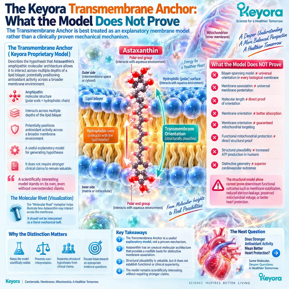
The Keyora Transmembrane Anchor: What the Model Does Not Prove
The Transmembrane Anchor is best treated as an explanatory membrane model rather than a clinically proven mechanical mechanism
Within the Keyora Cardiac Architecture, The Transmembrane Anchor can remain a useful proprietary framework.
But its definition should be evidence-bound.
A scientifically disciplined definition would be:
The Transmembrane Anchor describes the hypothesis that Astaxanthin’s amphipathic molecular architecture allows it to interact across multiple depths of a lipid bilayer, potentially positioning antioxidant activity across a broader membrane environment.
The related Molecular Rivet metaphor can also remain useful when it is clearly presented as a visualization.
It should not mean that Astaxanthin literally functions as a mechanical bolt.
This distinction matters because Keyora Astaxanthin EP-4 sometimes extends the structural model into stronger claims about membrane stabilization, electron-transport alignment, reduced electron leakage, and preservation of mitochondrial voltage.
Those functional hypotheses may be scientifically interesting.
But the structural model alone cannot prove them.
The evidence boundaries are:
-
Bilayer-spanning model ≠ universal orientation in every biological membrane
-
Membrane association ≠ universal membrane penetration
-
Molecular length ≠ direct proof of orientation
-
Membrane orientation ≠ better absorption
-
Membrane orientation ≠ guaranteed mitochondrial targeting
-
Functional mitochondrial protection ≠ direct structural proof
-
Structural plausibility ≠ increased ATP production in humans
-
Distinctive geometry ≠ superior cardiovascular outcomes
The most useful conclusion is therefore not that Astaxanthin is a literal membrane “steel beam.”
It is that Astaxanthin has an unusual molecular architecture that provides a credible mechanistic basis for distinctive membrane association.
That is enough to make the structure scientifically interesting.
It does not need to be turned into a stronger clinical claim.
This distinction also prepares the next question in the Keyora Cardiac Architecture:
Does Stronger Antioxidant Activity Mean Better Heart Protection?
This article is for educational and informational purposes only. It does not provide medical advice, diagnosis, treatment, cure, prevention, disease outcome claims, hormone restoration claims, fertility outcome claims, or formula-specific clinical efficacy claims.