
Can Astaxanthin Really Span the Lipid Bilayer?
Keyora Research Q&A Library
This is part of the Keyora Research Q&A Series, derived from Keyora Astaxanthin Research Journal Series.
Within the Keyora Astaxanthin Researcn framework, this Q&A translates complex astaxanthin biology into reader-friendly, evidence-bound answers, focusing on natural astaxanthin identity, molecular structure, antioxidant and redox mechanisms, membrane lipid interaction, mitochondrial resilience, inflammatory signaling pathways, human evidence interpretation, and the scientific principles behind responsible supplementation.
First published by Keyora Research Journal: www.keyorahealth.com

Direct Answer
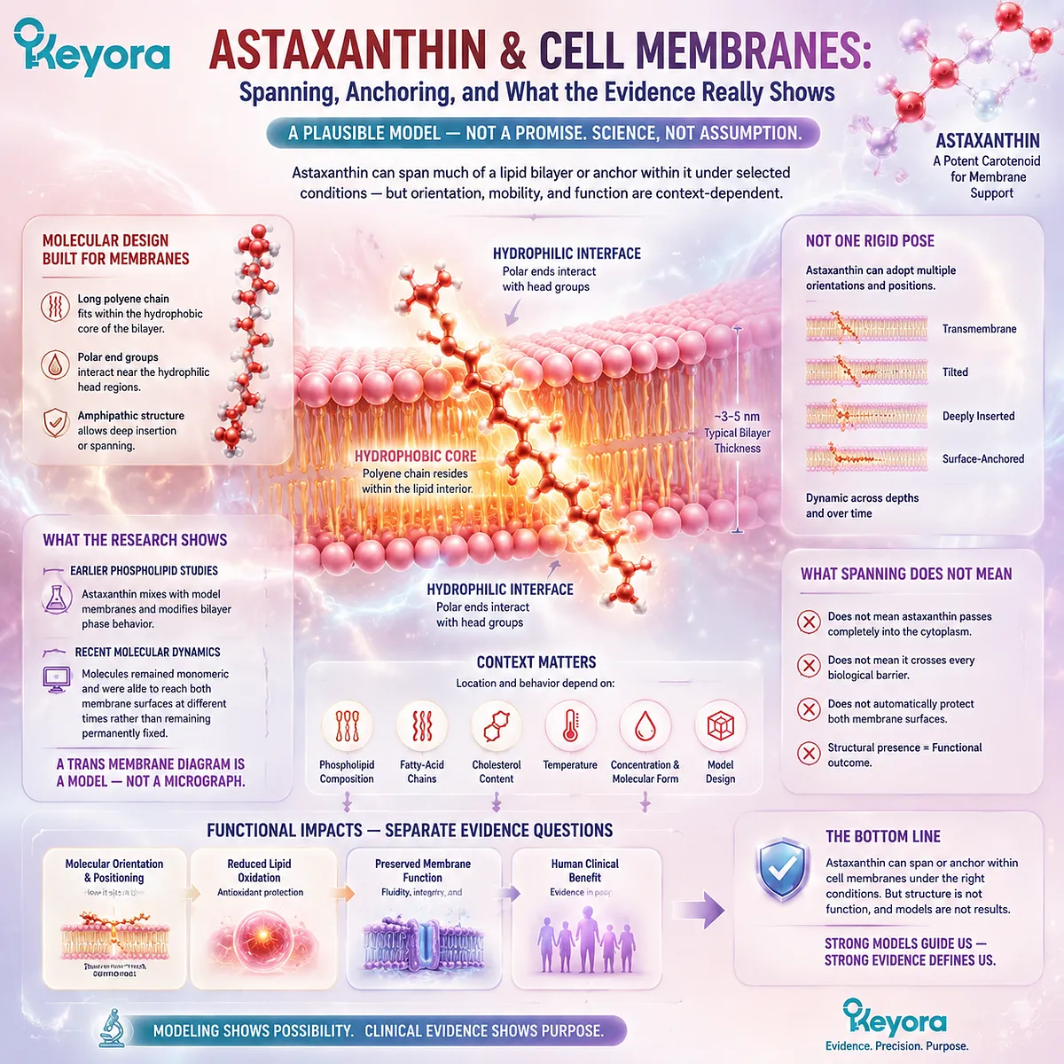
Astaxanthin can plausibly span much of a lipid bilayer or become strongly anchored within it under selected experimental conditions. Its long lipid-compatible central chain can occupy the membrane’s hydrophobic interior, while its oxygen-containing terminal regions can interact more readily near the polar phospholipid interfaces.
However, “membrane-spanning” should not be interpreted as one rigid pose adopted by every astaxanthin molecule. The molecule may be tilted, deeply inserted, associated more strongly with one interface, or mobile across different membrane depths. Its position can change with phospholipid composition, fatty-acid chains, cholesterol, temperature, concentration, molecular form, and model design.
Earlier phospholipid studies established that astaxanthin mixes with model membranes and changes bilayer phase behavior. More recent astaxanthin-specific molecular-dynamics research found that molecules remained monomeric in the modeled membrane and could reach both membrane surfaces at different times rather than remaining permanently fixed across both interfaces.
A transmembrane diagram therefore represents a plausible structural model, not a photograph of astaxanthin inside every human cell.
Spanning the bilayer also does not mean that astaxanthin passes completely through the membrane into the cytoplasm, crosses every biological barrier, or automatically protects both membrane surfaces. Molecular orientation, reduced lipid oxidation, preserved membrane function, and human clinical benefit are separate evidence questions.
What “Spanning the Lipid Bilayer” Actually Means
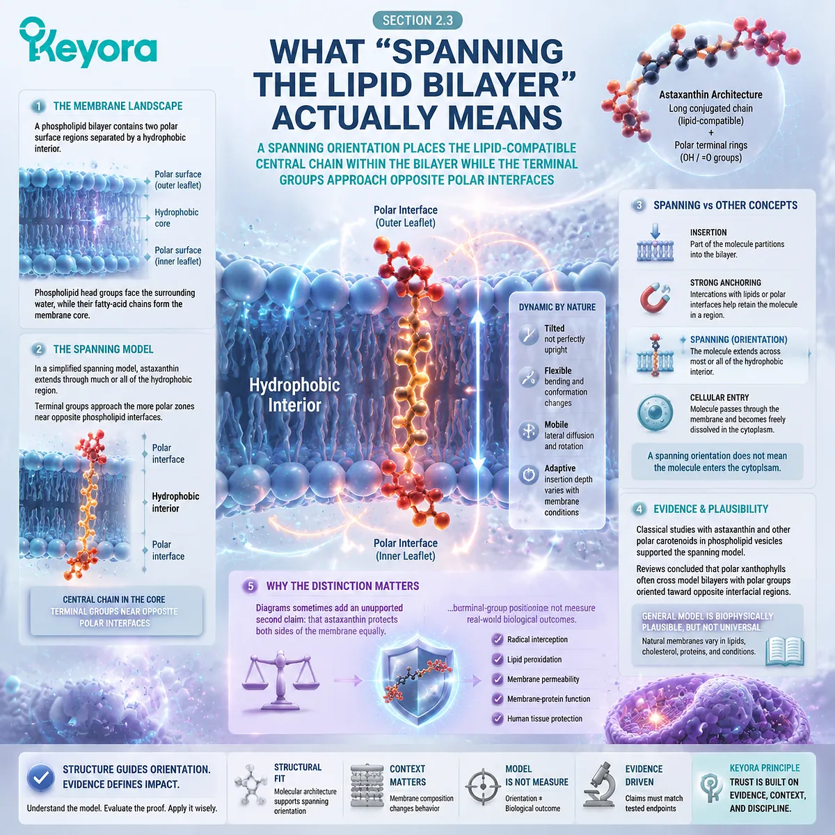
A spanning orientation places the lipid-compatible central chain within the bilayer while the terminal groups approach opposite polar interfaces
A phospholipid bilayer contains two polar surface regions separated by a hydrophobic interior. Phospholipid head groups face the surrounding water, while their fatty-acid chains form the membrane core.
Astaxanthin has a broadly corresponding molecular architecture. Its long conjugated chain is strongly compatible with lipid environments, while its two terminal rings contain hydroxyl and keto groups that create relatively polar regions.
In a simplified spanning model, the central chain extends through much or all of the membrane’s hydrophobic region. The terminal groups approach the more polar zones near opposite phospholipid interfaces.
The word “spanning” does not require the molecule to stand perfectly upright. A molecule can cross most of the hydrophobic region while remaining tilted relative to the membrane normal. It can also bend, rotate, move laterally, or change insertion depth as the surrounding lipids move.
This is different from merely entering a membrane. Insertion means that part of the molecule partitions into the bilayer. Strong anchoring means that interactions with lipids or polar interfaces help retain the molecule within a particular region. Spanning is a more specific orientation in which the molecule extends across most or all of the hydrophobic interior.
These terms are also different from cellular entry. An astaxanthin molecule can occupy a transmembrane orientation without passing through the membrane and becoming freely dissolved in the cytoplasm.
Classical research using astaxanthin and other polar carotenoids in phospholipid vesicles contributed to the spanning model. Later reviews concluded that polar xanthophylls often cross model bilayers with their polar groups oriented toward opposite interfacial regions. This general model remains biophysically plausible, but it does not establish identical behavior in every natural membrane.
The distinction matters because diagrams often add a second unsupported claim: that astaxanthin protects both sides of the membrane equally. Terminal-group positioning may support such a hypothesis, but orientation alone does not measure radical interception, lipid oxidation, permeability, membrane-protein function, or human tissue protection.
Why Astaxanthin Can Adopt More Than One Orientation
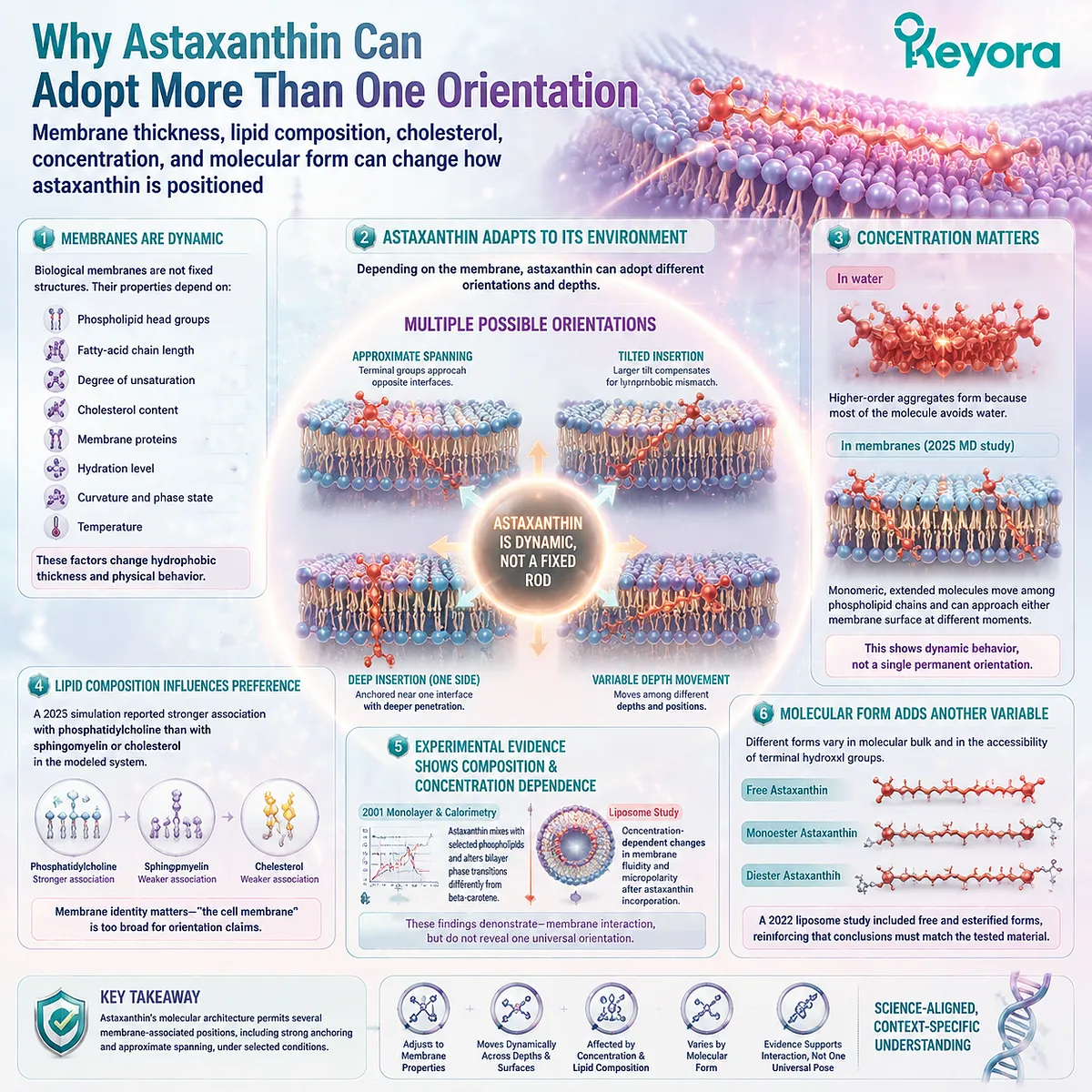
Membrane thickness, lipid composition, cholesterol, concentration, and molecular form can change how astaxanthin is positioned
A biological membrane is not a fixed geometric structure. Its hydrophobic thickness and physical behavior depend on phospholipid head groups, fatty-acid chain length, unsaturation, cholesterol, proteins, hydration, curvature, and temperature.
Astaxanthin must adjust to this changing environment. In one bilayer, its terminal regions may approach opposite interfaces. In another, hydrophobic mismatch may favor a larger tilt, deeper insertion from one side, or movement between different depths.
Concentration also matters. Astaxanthin can form aggregates in aqueous environments because most of the molecule avoids water. The 2025 molecular-dynamics study reported higher-order aggregation in water but monomeric astaxanthin after incorporation into its modeled complex membrane. The molecules remained extended, moved among phospholipid chains, and could approach either membrane surface at different moments.
That result does not prove that spanning models are false. It shows that the molecule may behave dynamically rather than functioning as a permanently fixed rod joining both interfaces at once.
Phospholipid identity may also affect preferred localization. The same 2025 simulation reported stronger association with phosphatidylcholine than with sphingomyelin or cholesterol in the modeled system. This is a model-specific observation, but it illustrates why “the cell membrane” is too broad a description for an orientation claim.
Experimental work also shows that astaxanthin changes membrane behavior according to composition and concentration. A 2001 monolayer and calorimetry study found that astaxanthin mixed with selected phospholipids and altered bilayer phase transitions differently from beta-carotene. A separate liposome study found concentration-dependent changes in membrane fluidity and micropolarity after astaxanthin incorporation.
These findings demonstrate membrane interaction, but neither phase-transition changes nor fluidity measurements directly reveal one universal orientation.
Molecular form adds another variable. Free astaxanthin, monoesters, and diesters differ in molecular bulk and in the accessibility of terminal hydroxyl groups. A 2022 liposome investigation included free and esterified astaxanthin forms, reinforcing the need to match conclusions to the tested material rather than assigning one membrane model to every commercial ingredient.
The safest conclusion is not that astaxanthin always stands vertically or always lies sideways. It is that its architecture permits several membrane-associated positions, including strong anchoring and approximate spanning under selected conditions.
What Membrane Models Can and Cannot Show
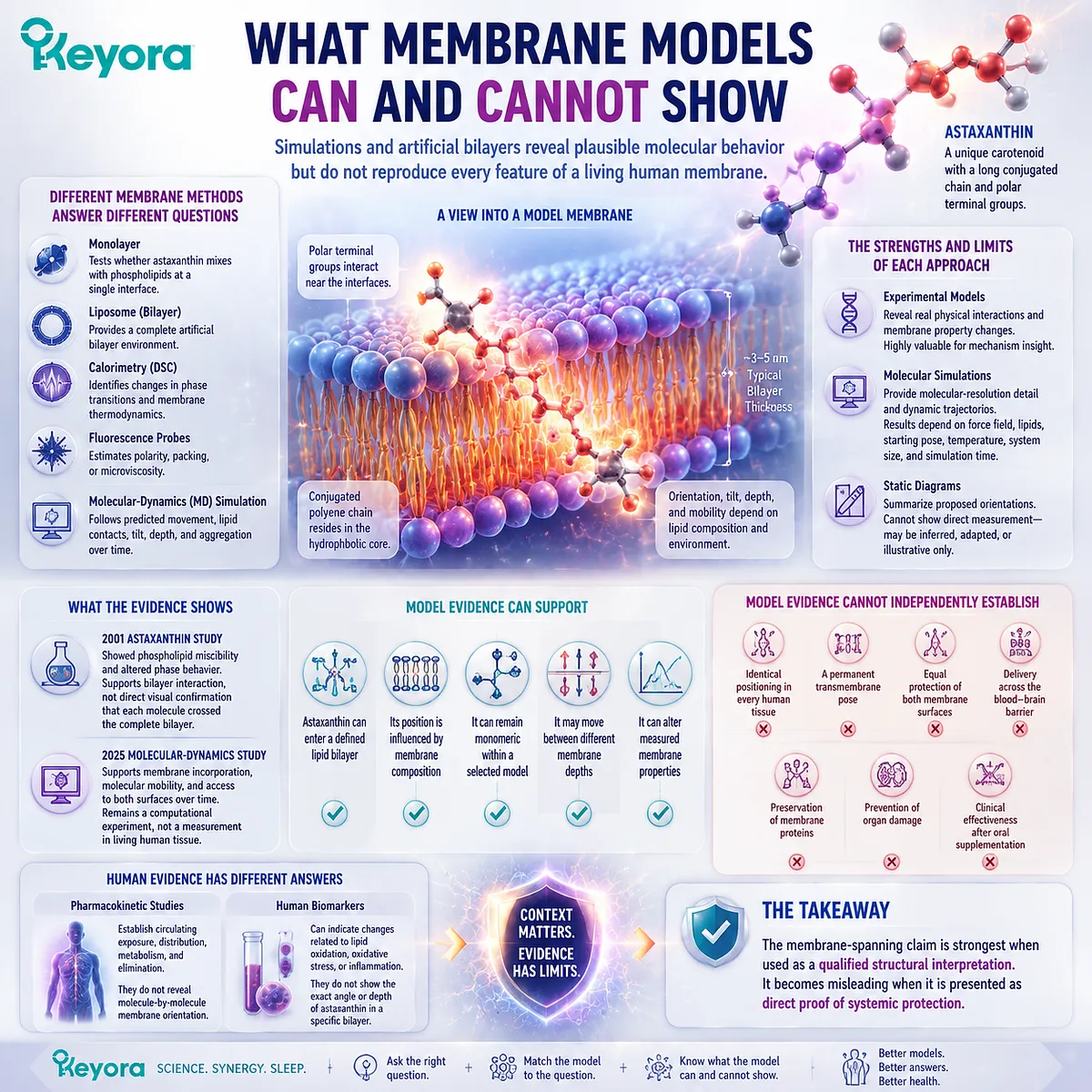
Simulations and artificial bilayers reveal plausible molecular behavior but do not reproduce every feature of a living human membrane
Different membrane methods answer different questions.
A monolayer can test whether astaxanthin mixes with phospholipids at a single interface. A liposome provides a complete artificial bilayer. Calorimetry can identify changes in phase transitions, while fluorescence probes can estimate polarity, packing, or microviscosity.
Molecular-dynamics simulations can follow predicted movement, lipid contacts, tilt, depth, and aggregation over time. Their advantage is molecular detail, but the result depends on the selected force field, lipid mixture, starting position, temperature, system size, and simulation duration.
A static diagram is one step further removed. It may summarize a proposed orientation, but it cannot show whether the arrangement was measured directly, inferred from membrane properties, borrowed from another xanthophyll, or created only for illustration.
The 2001 astaxanthin study showed phospholipid miscibility and altered phase behavior. Those findings support bilayer interaction, not direct visual confirmation that each molecule crossed the complete bilayer.
The 2025 simulation provides more detailed astaxanthin-specific information. It supports membrane incorporation, molecular mobility, and access to both surfaces over time, but it remains a computational experiment rather than a measurement in living human tissue.
Model evidence can support conclusions such as:
-
astaxanthin can enter a defined lipid bilayer
-
its position is influenced by membrane composition
-
it can remain monomeric within a selected model
-
it may move between different membrane depths
-
it can alter measured membrane properties
The same evidence cannot independently establish:
-
identical positioning in every human tissue
-
a permanent transmembrane pose
-
equal protection of both membrane surfaces
-
delivery across the blood-brain barrier
-
preservation of membrane proteins
-
prevention of organ damage
-
clinical effectiveness after oral supplementation
Human pharmacokinetic evidence would establish circulating exposure, not molecule-by-molecule membrane orientation. A human biomarker could indicate a change related to lipid oxidation, but it would not reveal the exact angle adopted by astaxanthin in a particular bilayer.
The membrane-spanning claim is therefore strongest when used as a qualified structural interpretation. It becomes misleading when it is presented as direct proof of systemic protection.
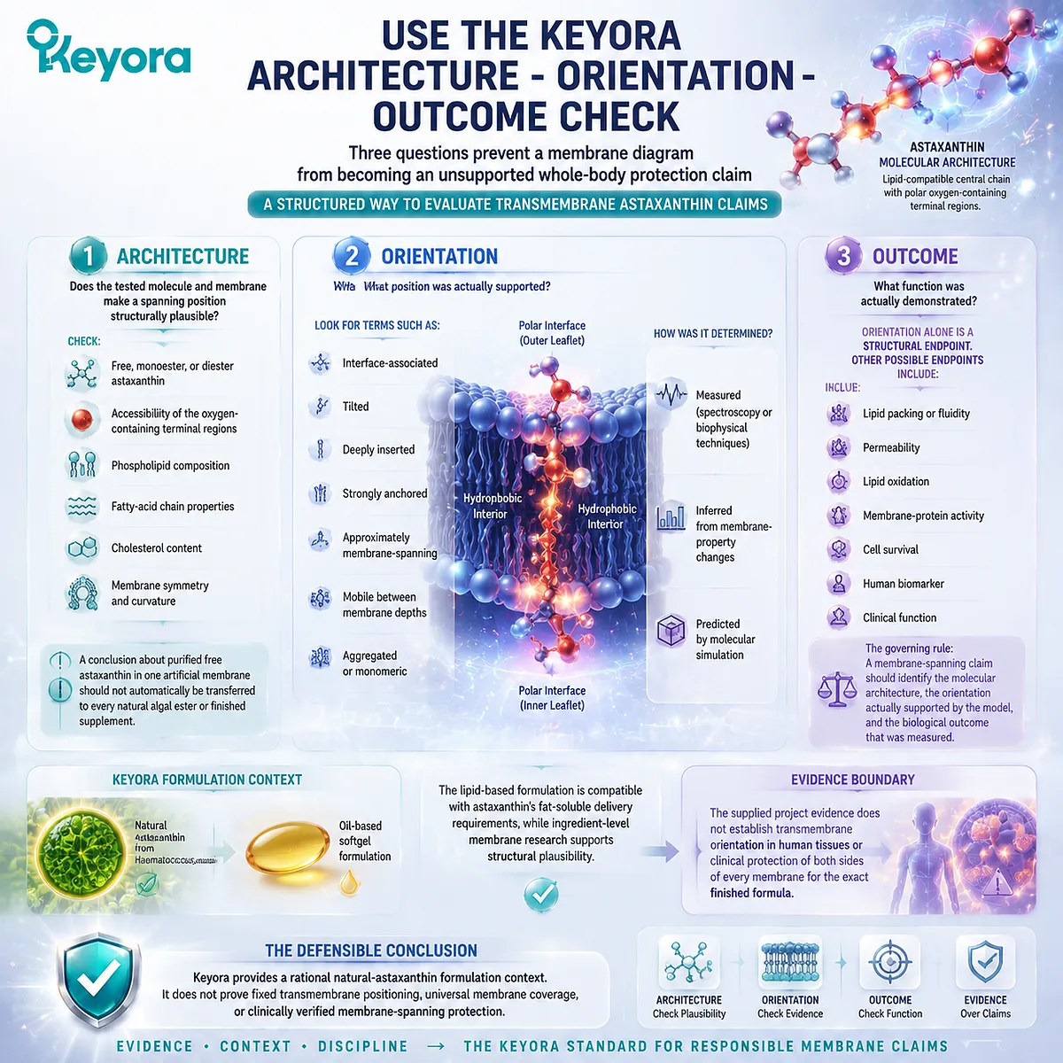
Use the Keyora Architecture – Orientation – Outcome Check
Three questions prevent a membrane diagram from becoming an unsupported whole-body protection claim
The Keyora Architecture – Orientation – Outcome Check provides a practical method for evaluating transmembrane astaxanthin claims.
1. Architecture
Does the tested molecule and membrane make a spanning position structurally plausible?
Check:
-
free, monoester, or diester astaxanthin
-
accessibility of the oxygen-containing terminal regions
-
phospholipid composition
-
fatty-acid chain properties
-
cholesterol content
-
membrane symmetry and curvature
A conclusion about purified free astaxanthin in one artificial membrane should not automatically be transferred to every natural algal ester or finished supplement.
2. Orientation
What position was actually supported?
Look for terms such as:
-
interface-associated
-
tilted
-
deeply inserted
-
strongly anchored
-
approximately membrane-spanning
-
mobile between membrane depths
-
aggregated or monomeric
Then identify how the position was obtained. Was it measured through spectroscopy or another biophysical technique, inferred from membrane-property changes, or predicted by molecular simulation?
3. Outcome
What function was actually demonstrated?
Orientation alone is a structural endpoint. Other possible endpoints include lipid packing, fluidity, permeability, lipid oxidation, membrane-protein activity, cell survival, a human biomarker, or a clinical function.
The governing rule is:
A membrane-spanning claim should identify the molecular architecture, the orientation actually supported by the model, and the biological outcome that was measured.
Keyora uses natural astaxanthin from Haematococcus pluvialis in an oil-based softgel context. The lipid-based formulation is compatible with astaxanthin’s fat-soluble delivery requirements, while ingredient-level membrane research supports structural plausibility. The supplied project evidence does not establish transmembrane orientation in human tissues or clinical protection of both sides of every membrane for the exact finished formula.
The defensible conclusion is that Keyora provides a rational natural-astaxanthin formulation context. It does not prove fixed transmembrane positioning, universal membrane coverage, or clinically verified membrane-spanning protection.
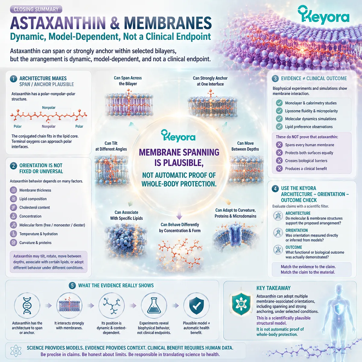
Closing Summary
Astaxanthin can span or strongly anchor within selected bilayers, but the arrangement is dynamic, model-dependent, and not a clinical endpoint
Astaxanthin’s polar – nonpolar – polar architecture makes a membrane-spanning or strongly anchored orientation plausible. Its conjugated central chain can occupy the lipid core, while its oxygen-containing terminal regions may approach polar membrane interfaces.
The position is not necessarily vertical, permanent, or identical in every membrane. Astaxanthin may tilt, rotate, move between depths, associate preferentially with certain lipids, or adopt different behavior according to concentration and molecular form.
Biophysical experiments and molecular simulations provide valuable evidence about membrane interaction. They do not prove that every molecule spans every human membrane, protects both surfaces equally, crosses biological barriers, or produces a clinical benefit.
Use the Keyora Architecture – Orientation – Outcome Check. Confirm that the molecular and membrane structures support the proposed arrangement, identify whether orientation was measured or inferred, and determine which functional outcome was actually demonstrated.
Membrane spanning is a scientifically plausible structural model. It is not automatic proof of whole-body protection.
This article is for educational and informational purposes only. It does not provide medical advice, diagnosis, treatment, cure, prevention, disease outcome claims, hormone restoration claims, fertility outcome claims, or formula-specific clinical efficacy claims.