
Can Astaxanthin Protect the Heart During Ischemia-Reperfusion Injury? What Does the Evidence Actually Show?
Keyora Research Q&A Library
This is part of the Keyora Research Q&A Series, derived from Keyora Astaxanthin Research Journal Series.
Within the Keyora Astaxanthin Researcn framework, this Q&A translates complex astaxanthin biology into reader-friendly, evidence-bound answers, focusing on natural astaxanthin identity, molecular structure, antioxidant and redox mechanisms, membrane lipid interaction, mitochondrial resilience, inflammatory signaling pathways, human evidence interpretation, and the scientific principles behind responsible supplementation.
First published by Keyora Research Journal: www.keyorahealth.com
Direct Answer
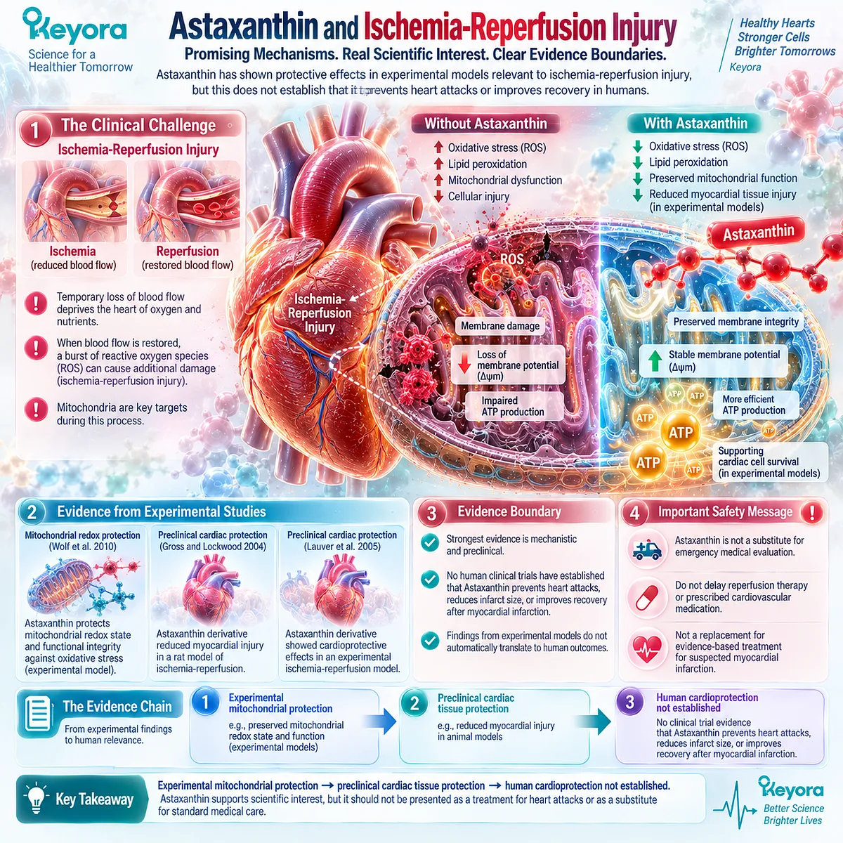
Astaxanthin has shown protective effects in experimental models relevant to ischemia-reperfusion injury, including effects on mitochondrial redox integrity and myocardial tissue injury.
However, these findings do not establish that Astaxanthin prevents heart attacks, reduces infarct size, or improves recovery after myocardial infarction in humans.
The strongest evidence discussed in Keyora Astaxanthin EP-4: The Cardiac Architecture: Bio-Energetic Sovereignty is mechanistic and preclinical.
Wolf et al. (2010), Astaxanthin Protects Mitochondrial Redox State and Functional Integrity Against Oxidative Stress, supports an experimental mitochondrial-protection mechanism.
Gross and Lockwood (2004) and Lauver et al. (2005) provide preclinical cardiac ischemia-reperfusion evidence involving an Astaxanthin derivative rather than a human trial of ordinary oral natural Astaxanthin.
The evidence therefore supports scientific interest, not a treatment claim.
The correct conclusion is:
Experimental mitochondrial protection → preclinical cardiac tissue protection → human cardioprotection not established
Astaxanthin supplementation should never be presented as a substitute for emergency evaluation, reperfusion therapy, prescribed cardiovascular medication, or other evidence-based treatment for suspected myocardial infarction.
What Is Ischemia-Reperfusion Injury?
Restoring blood flow is essential, but stressed tissue can experience additional oxidative and mitochondrial injury during reperfusion
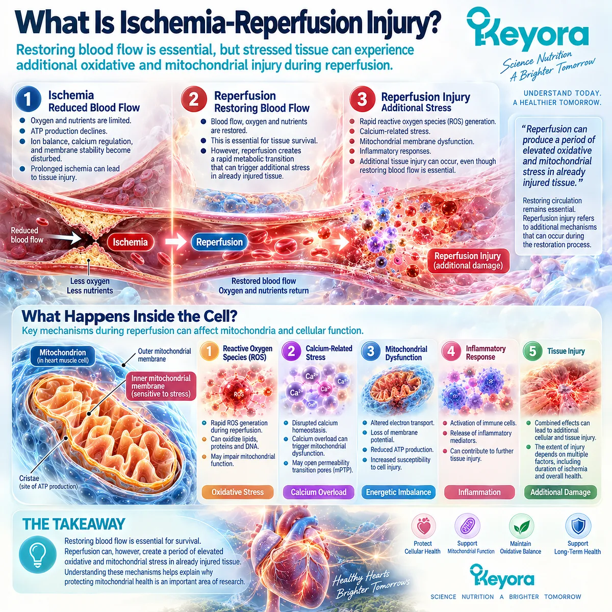
Ischemia occurs when blood flow to a tissue becomes severely restricted, reducing the supply of oxygen and metabolic substrates.
In heart muscle, prolonged ischemia threatens ATP production because oxidative phosphorylation depends on oxygen availability. As energy supply becomes inadequate, ion handling, calcium regulation, membrane stability, and multiple cellular processes can become progressively disturbed.
Reperfusion means restoring blood flow.
This is essential because ischemic tissue needs oxygen and nutrients to survive. However, reperfusion can also create a period of intense metabolic transition.
Mitochondria that were operating under oxygen-restricted conditions are suddenly exposed to renewed oxygen availability while electron transport, calcium balance, pH, and cellular redox systems may still be disrupted.
This can contribute to rapid reactive oxygen species generation, calcium-related stress, mitochondrial membrane dysfunction, and additional tissue injury.
In Keyora Astaxanthin EP-4, this transition is described with terms such as the “Reperfusion Blast.” The underlying biological concept is legitimate, but the public scientific interpretation should remain more precise: reperfusion can produce a period of elevated oxidative and mitochondrial stress in already injured tissue.
Reperfusion injury is therefore not evidence that restoring blood flow is harmful overall.
Restoring circulation remains essential.
The concept refers to additional injury mechanisms that can occur during the restoration process.
Why Mitochondria Matter During Reperfusion
Mitochondrial membrane potential, calcium handling, redox balance, and permeability regulation become important during severe reperfusion stress
Mitochondria sit near the center of ischemia-reperfusion biology because several stress pathways converge on them.
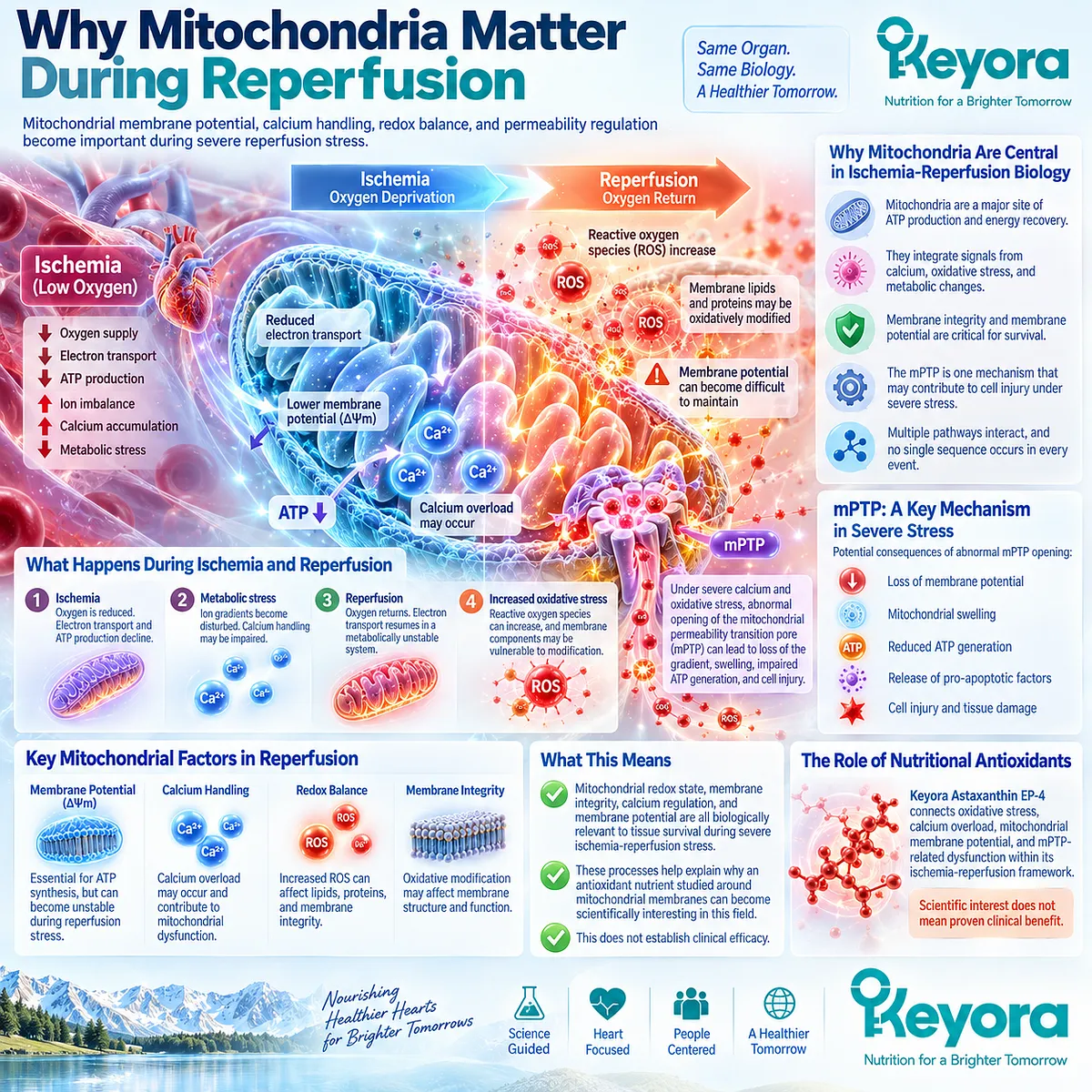
During ischemia, reduced oxygen availability limits normal electron transport and ATP production.
As ATP availability declines, cells can become less able to maintain normal ion gradients. Calcium handling may become disturbed, and mitochondrial calcium burden can rise under severe stress.
When oxygen is restored, electron transport resumes in a system that may already be metabolically unstable.
Reactive oxygen species can increase.
Membrane lipids and proteins may be exposed to oxidative modification.
Mitochondrial membrane potential can become difficult to maintain.
One mechanism implicated in severe mitochondrial injury is the mitochondrial permeability transition pore, commonly abbreviated mPTP.
Under major calcium and oxidative stress, abnormal permeability transition can contribute to loss of the electrochemical gradient, swelling, impaired ATP generation, and cell injury.
Keyora Astaxanthin EP-4 connects oxidative stress, calcium overload, mitochondrial membrane potential, and mPTP-related dysfunction within its ischemia-reperfusion framework.
But these processes should not be presented as one guaranteed sequence occurring identically in every ischemic event.
Ischemia-reperfusion injury involves multiple interacting pathways.
The important conclusion is narrower:
Mitochondrial redox state, membrane integrity, calcium regulation, and membrane potential are all biologically relevant to tissue survival during severe ischemia-reperfusion stress.
This explains why an antioxidant nutrient studied around mitochondrial membranes can become scientifically interesting in this field.
It does not establish clinical efficacy.
What the Mitochondrial Evidence Shows
Wolf et al. supports an experimental mitochondrial-protection mechanism, not a human cardiac outcome
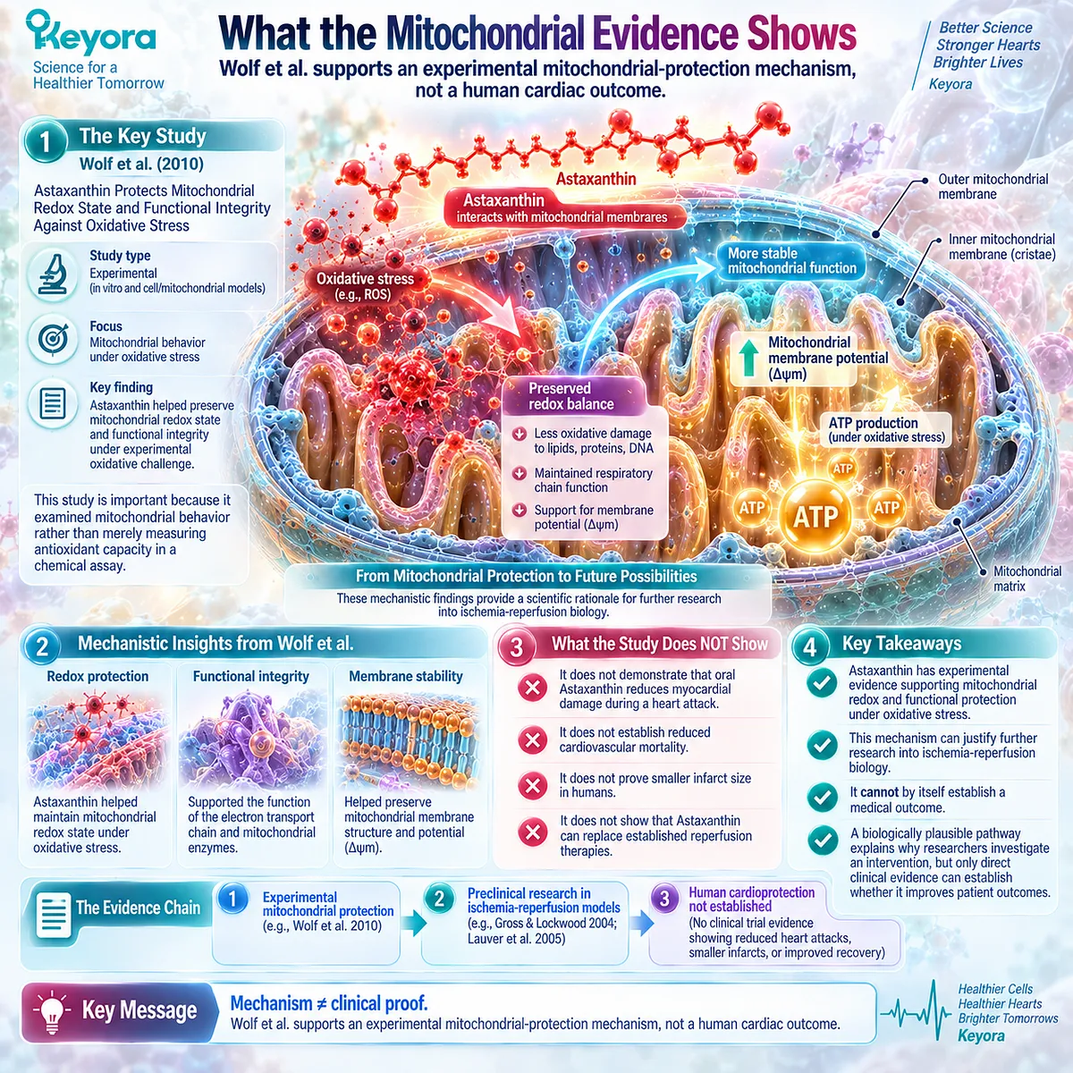
One of the central mechanistic studies is Wolf et al. (2010), Astaxanthin Protects Mitochondrial Redox State and Functional Integrity Against Oxidative Stress.
This study is important because it examined mitochondrial behavior under oxidative stress rather than merely measuring antioxidant capacity in a chemical assay.
In Keyora Astaxanthin EP-4: The Cardiac Architecture: Bio-Energetic Sovereignty, the Wolf study is used to support the hypothesis that Astaxanthin can help preserve mitochondrial redox state and functional integrity under experimental oxidative challenge. The article also connects these findings with membrane potential and mitochondrial stress resistance.
That is meaningful mechanistic evidence.
But the evidence level must remain clear.
Wolf et al. does not demonstrate that a person taking oral Astaxanthin will experience less myocardial damage during a heart attack.
It does not establish reduced cardiovascular mortality.
It does not prove smaller infarct size in humans.
It does not show that Astaxanthin can replace established reperfusion therapies.
The appropriate interpretation is:
Astaxanthin has experimental evidence supporting mitochondrial redox and functional protection under oxidative stress.
That mechanism can justify further research into ischemia-reperfusion biology.
It cannot by itself establish a medical outcome.
This distinction reflects one of the most important rules in evidence interpretation:
Mechanism ≠ clinical proof
A biologically plausible pathway can explain why researchers investigate an intervention.
Only direct clinical evidence can establish whether that intervention improves patient outcomes.
What the Cardiac Injury Studies Actually Show
Preclinical Astaxanthin-derivative studies report smaller myocardial injury, but they are not human prevention trials
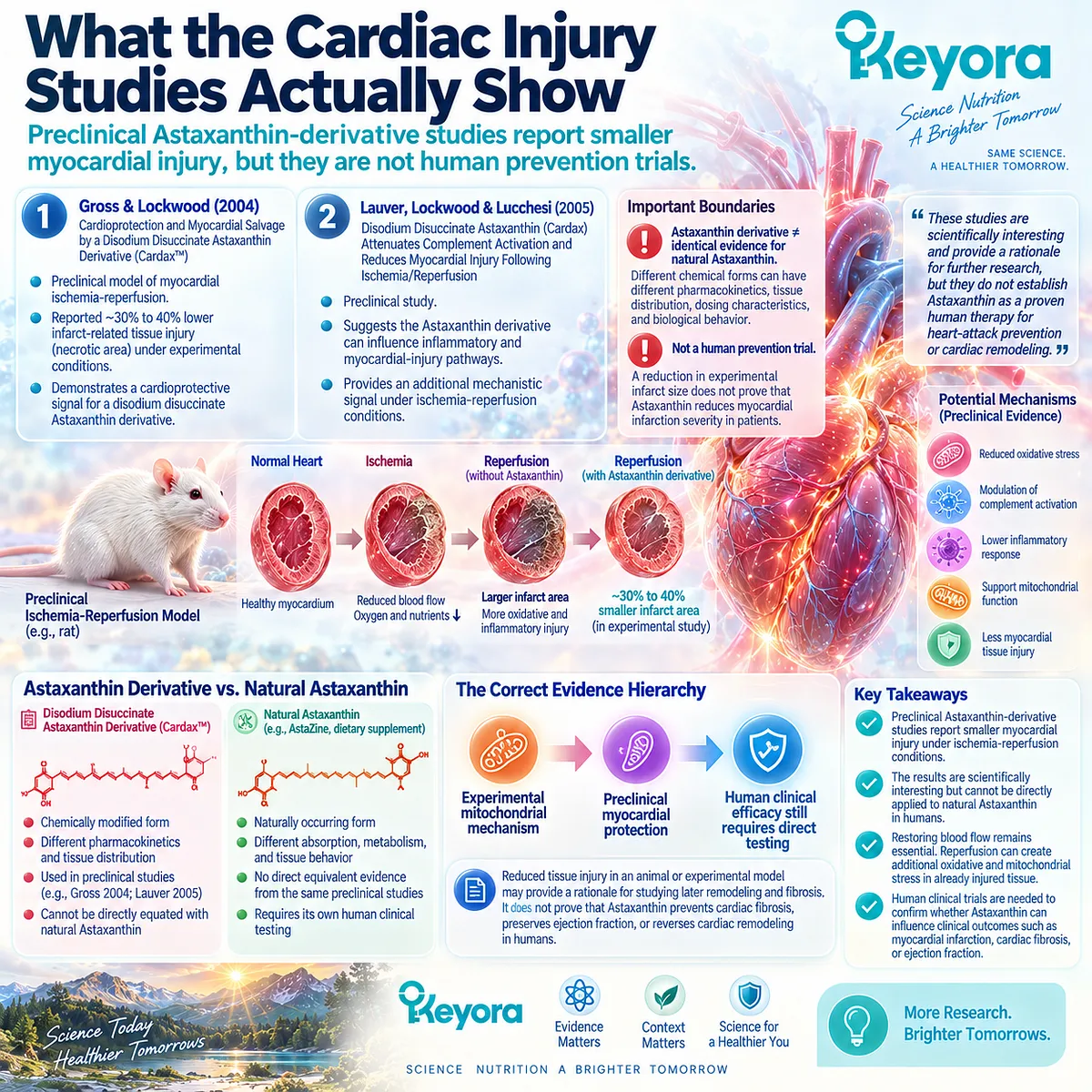
The most striking cardiac evidence discussed in Keyora Astaxanthin EP-4 comes from Gross and Lockwood (2004), Cardioprotection and Myocardial Salvage by a Disodium Disuccinate Astaxanthin Derivative (Cardax™).
The study examined myocardial ischemia-reperfusion in a preclinical model and reported a reduction in infarct-related tissue injury. Keyora Astaxanthin EP-4 describes necrotic area as being approximately 30% to 40% lower under the experimental conditions discussed.
This result is scientifically interesting.
But two boundaries are essential.
First, the intervention was a disodium disuccinate Astaxanthin derivative, not simply ordinary dietary natural Astaxanthin.
That matters because:
Astaxanthin derivative ≠ identical evidence for natural Astaxanthin
Different chemical forms can have different pharmacokinetics, tissue distribution, dosing characteristics, and biological behavior.
Therefore, the result cannot be directly transferred to oral natural Astaxanthin, AstaZine, or a finished consumer supplement.
Second, this was not a human heart-attack prevention trial.
A reduction in experimental infarct size is not equivalent to proving that Astaxanthin reduces myocardial infarction severity in patients.
The same evidence boundary applies to Lauver, Lockwood, and Lucchesi (2005), Disodium Disuccinate Astaxanthin (Cardax) Attenuates Complement Activation and Reduces Myocardial Injury Following Ischemia/Reperfusion.
This study adds another preclinical signal suggesting that an Astaxanthin derivative can influence inflammatory and myocardial-injury pathways under ischemia-reperfusion conditions. It does not convert the evidence into established human therapy.
The correct evidence hierarchy is:
Experimental mitochondrial mechanism
→ preclinical myocardial protection
→ human clinical efficacy still requires direct testing
This hierarchy also prevents another overinterpretation.
Reduced tissue injury in an animal or experimental model may provide a rationale for studying later remodeling and fibrosis.
It does not prove that Astaxanthin prevents cardiac fibrosis, preserves ejection fraction, or reverses cardiac remodeling in humans.
The Evidence Verdict: Promising Mechanism, Unproven Human Protection
Astaxanthin remains scientifically interesting for reperfusion biology, but it should not be presented as a treatment or preventive strategy for heart attack
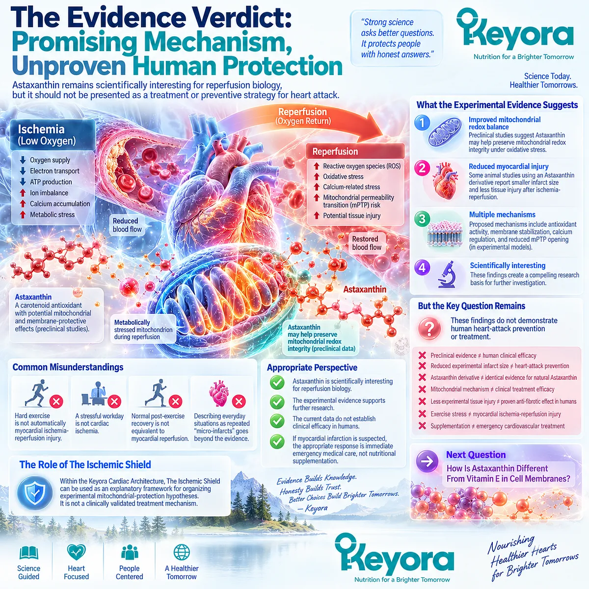
The ischemia-reperfusion evidence creates a compelling biological research story.
Ischemia restricts oxygen and disrupts energy metabolism.
Reperfusion restores blood flow but can expose metabolically stressed mitochondria to additional oxidative and calcium-related challenges.
Experimental Astaxanthin research suggests that mitochondrial redox integrity may be better preserved under oxidative stress.
Preclinical studies using an Astaxanthin derivative also report reductions in myocardial injury under ischemia-reperfusion conditions.
But the final step has not been established:
These findings do not demonstrate human heart-attack prevention or treatment.
That distinction is especially important because Keyora Astaxanthin EP-4 sometimes extends preclinical observations into stronger descriptions of tissue preservation, fibrosis prevention, and long-term cardiac resilience.
The underlying experiments justify scientific investigation, but they do not justify those outcomes as established human clinical effects.
The same caution applies to ordinary exercise and stress.
Hard exercise is not automatically myocardial ischemia-reperfusion injury.
A stressful workday is not cardiac ischemia.
Normal post-exercise recovery is not equivalent to myocardial reperfusion.
And describing these everyday situations as repeated “micro-infarcts” or “micro-reperfusion injury” would go beyond the available evidence.
Within the Keyora Cardiac Architecture, The Ischemic Shield can therefore be used only as an explanatory framework for organizing experimental mitochondrial-protection hypotheses.
It is not a clinically validated treatment mechanism.
The evidence boundaries are:
-
Preclinical cardioprotection ≠ human cardioprotection
-
Reduced experimental infarct size ≠ heart-attack prevention
-
Astaxanthin derivative ≠ identical evidence for natural Astaxanthin
-
Mitochondrial mechanism ≠ clinical treatment efficacy
-
Less experimental tissue injury ≠ proven anti-fibrotic effect in humans
-
Exercise stress ≠ myocardial ischemia-reperfusion injury
-
Supplementation ≠ emergency cardiovascular treatment
Astaxanthin remains scientifically interesting because the experimental evidence identifies plausible mitochondrial and redox pathways worthy of further study.
But if myocardial infarction is suspected, the appropriate response is immediate emergency medical care, not nutritional supplementation.
With the oxidative-stress and mitochondrial-protection group now established, the next question moves into a different comparison:
How Is Astaxanthin Different From Vitamin E in Cell Membranes?
This article is for educational and informational purposes only. It does not provide medical advice, diagnosis, treatment, cure, prevention, disease outcome claims, hormone restoration claims, fertility outcome claims, or formula-specific clinical efficacy claims.