
Can Astaxanthin Support Myelin and Nerve Health?
Keyora Research Q&A Library
This is part of the Keyora Research Q&A Series, derived from Keyora Astaxanthin Research Journal Series.
Within the Keyora Astaxanthin Researcn framework, this Q&A translates complex astaxanthin biology into reader-friendly, evidence-bound answers, focusing on natural astaxanthin identity, molecular structure, antioxidant and redox mechanisms, membrane lipid interaction, mitochondrial resilience, inflammatory signaling pathways, human evidence interpretation, and the scientific principles behind responsible supplementation.
First published by Keyora Research Journal: www.keyorahealth.com
Direct Answer
Yes
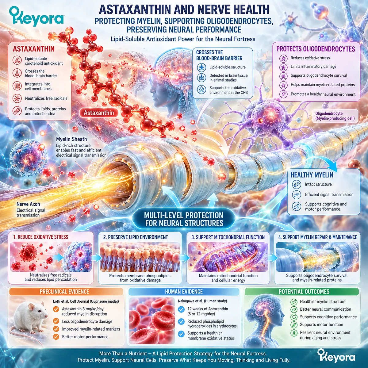
Astaxanthin has promising preclinical evidence for supporting myelin related structures and protecting oligodendrocytes under experimental injury conditions.
Its lipid compatible antioxidant properties provide an additional biological rationale for supporting the oxidative environment surrounding nerve cells.
Myelin is a specialized, lipid rich structure that wraps around nerve fibers and enables efficient electrical signal transmission. Its integrity depends not only on lipid composition but also on the health of the cells responsible for producing and maintaining it.
In a study published in Cell Journal, Lotfi and colleagues investigated Astaxanthin in a cuprizone induced rat model of demyelination.
Animals receiving 3 mg/kg/day Astaxanthin showed less myelin disruption and oligodendrocyte damage than untreated injury model groups. The researchers also observed favorable changes in myelin associated molecular markers and motor performance.
Separate human research provides another relevant evidence layer.
Nakagawa and colleagues demonstrated that 12 weeks of oral Astaxanthin supplementation at 6 or 12 mg/day reduced phospholipid hydroperoxides in human erythrocytes.
These findings support two connected but distinct conclusions:
-
Preclinical evidence – Astaxanthin has demonstrated protective effects on myelin related structures
-
Human evidence – Astaxanthin can influence measurable cellular membrane oxidation
This distinction develops the structural protection theme introduced in Keyora Astaxanthin EP-5: The Neural Fortress: A Mechanistic Analysis of Astaxanthin in Lipidomics Re-engineering and Neural Oxidative Debt.
The central concept is that supporting nerve health involves more than supplying nutrients.
The lipid environment and the cells maintaining neural structures must also remain biologically resilient.
What Is Myelin and Why Does Nerve Insulation Matter?
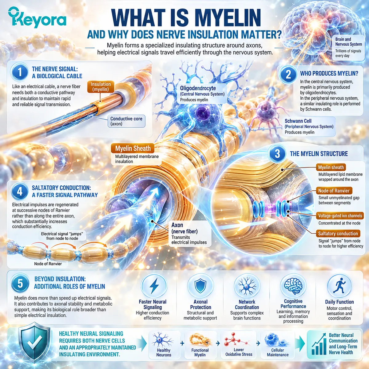
Myelin forms a specialized insulating structure around axons, helping electrical signals travel efficiently through the nervous system
Imagine sending an electrical signal along a long cable.
The cable needs both a conductive pathway and appropriate insulation. Without insulation, maintaining rapid and reliable transmission becomes considerably more difficult.
Myelin performs a comparable biological task.
It is a multilayered membrane structure wrapped around axons, the elongated nerve fibers responsible for transmitting electrical impulses between neurons and other target cells.
In the central nervous system, myelin is primarily produced by specialized cells called oligodendrocytes. In the peripheral nervous system, a similar insulating role is performed by Schwann cells.
The myelin sheath does not cover an axon continuously. Small exposed intervals called nodes of Ranvier separate successive myelinated segments.
These nodes contain specialized concentrations of voltage gated ion channels.
The arrangement enables saltatory conduction, in which electrical impulses are regenerated at successive nodes rather than requiring continuous regeneration along every part of the axonal membrane.
This architecture substantially increases conduction efficiency.
Myelin also contributes to axonal stability and metabolic support, making its biological role broader than simple electrical insulation.
For someone interested in long term nerve health, this creates an important distinction:
Healthy neural signaling requires both functioning nerve cells and an appropriately maintained insulating environment.
That is why nutritional research involving myelin considers not only neurons themselves but also oligodendrocytes, membrane composition, oxidative stress, and cellular maintenance.
Why Is Myelin So Rich in Lipids?
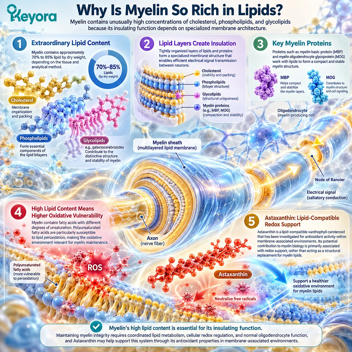
Myelin contains unusually high concentrations of cholesterol, phospholipids, and glycolipids because its insulating function depends on specialized membrane architecture
Myelin differs considerably from an ordinary cellular membrane.
Its tightly organized layers contain approximately 70% to 85% lipid by dry weight, depending on the tissue and analytical method.
Major lipid components include cholesterol, phospholipids, and specialized glycolipids such as galactocerebrosides.
These molecules are not interchangeable.
Cholesterol contributes to membrane organization and packing. Phospholipids form essential components of the lipid bilayers, while glycolipids participate in the distinctive structure and stability of myelin.
Proteins such as myelin basic protein (MBP) and myelin oligodendrocyte glycoprotein (MOG) also contribute to myelin biology, although they perform different structural and biological tasks.
This organization creates the insulating properties that make efficient nerve transmission possible.
However, the high lipid content also makes the surrounding oxidative environment relevant.
Biological membranes contain fatty acids with different degrees of unsaturation. Polyunsaturated fatty acids are particularly susceptible to lipid peroxidation because of their molecular structure.
Myelin maintenance therefore requires coordinated lipid metabolism, cellular redox regulation, and normal oligodendrocyte function.
This is where Astaxanthin becomes scientifically interesting.
As a lipid compatible xanthophyll carotenoid, Astaxanthin has been investigated for antioxidant activity within membrane associated environments.
Its potential contribution is primarily associated with redox support, rather than acting as a structural replacement for myelin lipids.
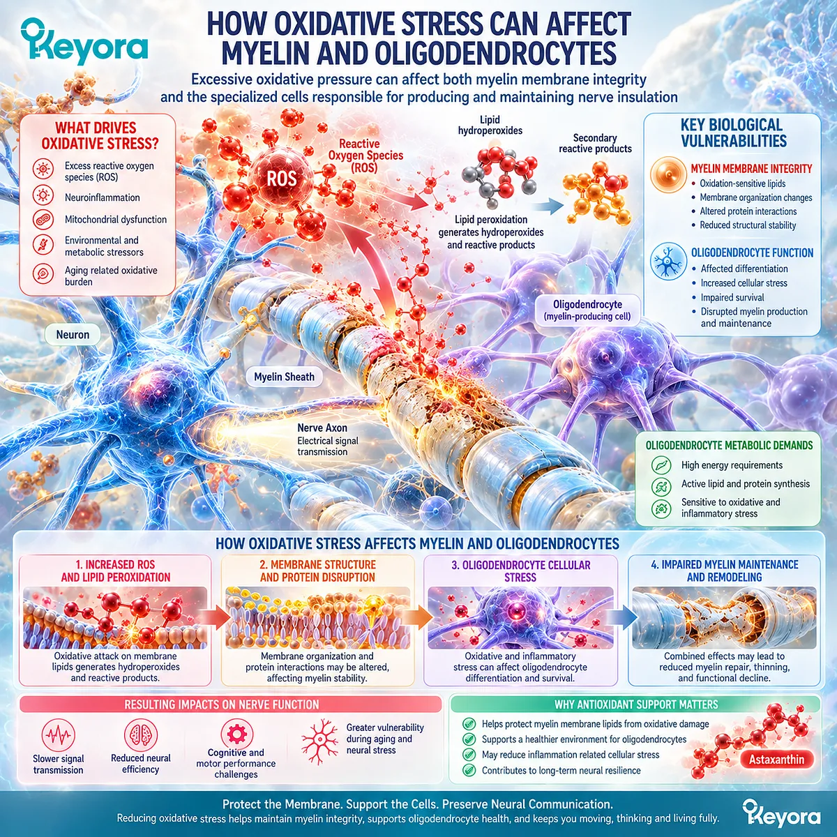
How Oxidative Stress Can Affect Myelin and Oligodendrocytes
Excessive oxidative pressure can affect both myelin membrane integrity and the specialized cells responsible for producing and maintaining nerve insulation
Myelin is not a structure that remains unchanged throughout life.
Its maintenance depends on ongoing cellular processes involving oligodendrocytes, lipid metabolism, protein synthesis, and the surrounding neural environment.
Oxidative stress can interfere with several of these processes.
When oxidation sensitive membrane lipids undergo lipid peroxidation, they may generate lipid hydroperoxides and secondary reactive products capable of affecting nearby cellular components.
Excessive oxidative modification can influence membrane organization and interactions involving proteins essential for normal cellular function.
Oligodendrocytes are another important consideration.
These specialized cells have substantial metabolic requirements associated with producing and maintaining myelin membranes. Their differentiation and survival can be affected by oxidative and inflammatory stress under certain experimental conditions.
This creates two related biological vulnerabilities:
Myelin Membrane Integrity
The lipid rich insulating structure itself may be affected by excessive oxidative reactions.
Oligodendrocyte Function
The cells responsible for myelin production and maintenance may experience cellular stress that interferes with their normal activity.
Protecting an existing membrane and preserving the cells responsible for maintaining that membrane are therefore different but complementary biological tasks.
Astaxanthin has attracted research interest because its antioxidant and anti-inflammatory activities may influence these processes in experimental neural injury models.
The important question is whether those mechanisms translate into measurable structural outcomes.
That is precisely what direct myelin related animal studies have begun to investigate.
What Astaxanthin Research Actually Shows About Myelin
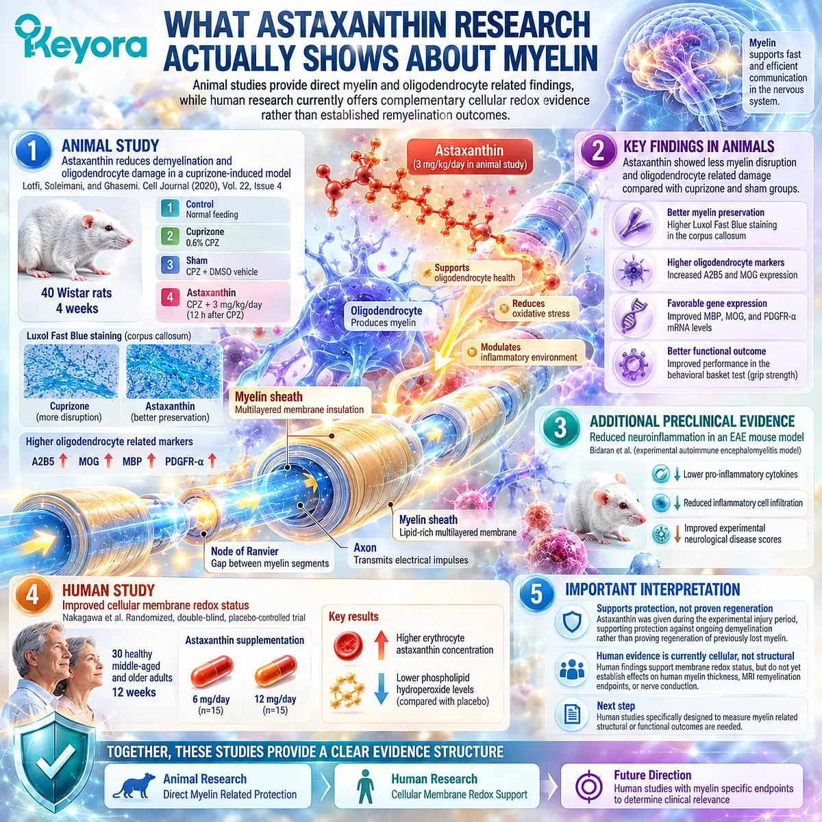
Animal studies provide direct myelin and oligodendrocyte related findings, while human research currently offers complementary cellular redox evidence rather than established remyelination outcomes
One of the most relevant studies was conducted by Lotfi, Soleimani, and Ghasemi and published in Cell Journal, Volume 22, Issue 4.
The researchers investigated whether Astaxanthin could reduce demyelination and oligodendrocyte damage in a cuprizone induced rat model.
Forty Wistar rats were randomly assigned to four experimental groups:
-
Control – Normal feeding
-
Cuprizone – 0.6% CPZ
-
Sham – CPZ plus DMSO vehicle
-
Astaxanthin – CPZ plus 3 mg/kg/day Astaxanthin
The experimental period lasted four weeks. Astaxanthin was administered 12 hours after the daily cuprizone exposure.
Researchers evaluated myelin integrity using Luxol Fast Blue staining, with particular attention to the corpus callosum.
They also investigated oligodendrocyte related proteins and genes, including A2B5, MOG, MBP, and PDGFR-alpha.
Compared with the cuprizone and sham groups, Astaxanthin treated animals showed less myelin disruption and oligodendrocyte related damage.
Myelin staining demonstrated better preservation of corpus callosum structure. Immunohistochemical findings showed higher expression of A2B5 and MOG markers, while molecular analysis identified favorable changes in MBP, MOG, and PDGFR-alpha gene expression.
The Astaxanthin group also performed better in the behavioral basket test, which assessed the animals’ ability to maintain their grip.
These findings are particularly valuable because they extend beyond measuring a general antioxidant biomarker. The study investigated actual myelin associated tissue changes alongside cellular markers and an animal functional endpoint.
However, Astaxanthin was administered during the experimental injury period. The findings therefore support protection against ongoing demyelinating injury rather than proving regeneration of previously lost human myelin.
Additional preclinical research provides supporting context.
Bidaran and colleagues examined Astaxanthin in an experimental autoimmune encephalomyelitis mouse model.
They observed reduced pro-inflammatory cytokine levels, lower inflammatory cell infiltration within central nervous system tissue, and favorable changes in experimental neurological disease scores.
This study contributes evidence about the inflammatory environment associated with neural injury, although it does not replace direct human myelin research.
Human findings provide a separate evidence layer.
Nakagawa and colleagues conducted a randomized, double-blind, placebo-controlled trial involving 30 healthy middle-aged and older adults.
After 12 weeks of supplementation with 6 or 12 mg Astaxanthin daily, erythrocyte Astaxanthin concentrations increased while phospholipid hydroperoxide levels were lower than in placebo.
This supports Astaxanthin’s relevance to human cellular membrane redox status.
It does not establish an effect on human myelin thickness, MRI remyelination endpoints, or restored nerve conduction.
Together, these studies provide a coherent evidence structure:
-
Animal Research – Direct Myelin Related Protection
-
Human Research – Cellular Membrane Redox Support
The next clinical step would require human studies specifically designed to measure myelin related structural or functional outcomes.
Where Keyora Asta 16MG Fits Into Nerve Health Support
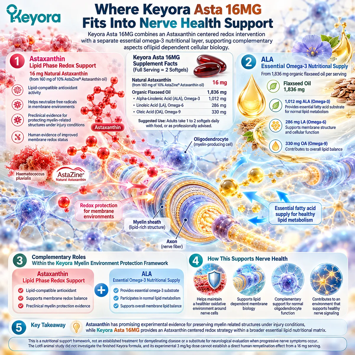
Keyora Asta 16MG combines an Astaxanthin centered redox intervention with a separate essential omega-3 nutritional layer, supporting complementary aspects of lipid dependent cellular biology
The current Keyora Asta 16MG Supplement Facts define a full serving as two softgels.
That serving provides:
16 mg Natural Astaxanthin, supplied by 160 mg of 10% AstaZine® Astaxanthin oil derived from Haematococcus pluvialis.
The formula also contains 1,836 mg organic flaxseed oil, including:
-
1,012 mg Alpha-Linolenic Acid (ALA), Omega-3
-
286 mg Linoleic Acid (LA), Omega-6
-
330 mg Oleic Acid (OA), Omega-9
The suggested adult use is one to two softgels daily with food, or as professionally advised.
For myelin and nerve health, Natural Astaxanthin remains the intervention protagonist.
Its relevance comes from lipid compatible antioxidant activity, direct preclinical myelin protection findings, and separate human evidence of improved membrane redox status.
ALA contributes a different nutritional task.
As an essential omega-3 fatty acid, ALA participates in normal lipid metabolism and provides dietary fatty acid substrate. It should not be treated as nutritionally identical to preformed DHA or described as clinically proven to regenerate human myelin.
Within the Keyora Myelin Environment Protection Framework, these roles remain distinct:
Astaxanthin – Lipid Phase Redox Support
ALA – Essential Omega-3 Nutritional Supply
Together, they provide a biologically complementary nutritional architecture rather than two ingredients performing the same function.
The Lotfi animal study did not investigate the finished Keyora formula, and its experimental 3 mg/kg dose cannot establish a direct human remyelination effect from a 16 mg serving.
The practical conclusion is therefore clear:
Astaxanthin has promising experimental evidence for preserving myelin related structures under injury conditions, while Keyora Asta 16MG provides an Astaxanthin centered redox strategy within a broader essential lipid nutritional matrix.
This is a nutritional support framework, not an established treatment for demyelinating disease or a substitute for neurological evaluation when progressive nerve symptoms occur.
This article is for educational and informational purposes only. It does not provide medical advice, diagnosis, treatment, cure, prevention, disease outcome claims, hormone restoration claims, fertility outcome claims, or formula-specific clinical efficacy claims.