
Can Astaxanthin Protect Brain Cell Membranes From Oxidative Stress?
Keyora Research Q&A Library
This is part of the Keyora Research Q&A Series, derived from Keyora Astaxanthin Research Journal Series.
Within the Keyora Astaxanthin Researcn framework, this Q&A translates complex astaxanthin biology into reader-friendly, evidence-bound answers, focusing on natural astaxanthin identity, molecular structure, antioxidant and redox mechanisms, membrane lipid interaction, mitochondrial resilience, inflammatory signaling pathways, human evidence interpretation, and the scientific principles behind responsible supplementation.
First published by Keyora Research Journal: www.keyorahealth.com
Direct Answer
Yes
Astaxanthin has meaningful scientific evidence supporting its role in lipid membrane oxidative protection, including a randomized human trial demonstrating reduced phospholipid hydroperoxides in erythrocytes after oral supplementation.
Its molecular structure also provides a biologically relevant explanation for why Astaxanthin is studied in the context of oxidation-sensitive neural membranes.
Brain cell membranes are not passive barriers.
They contain organized lipid structures that support ion transport, receptor activity, neurotransmission, mitochondrial function, and communication between neurons.
Their polyunsaturated fatty acid components are particularly important because they contribute to membrane properties while remaining susceptible to lipid peroxidation.
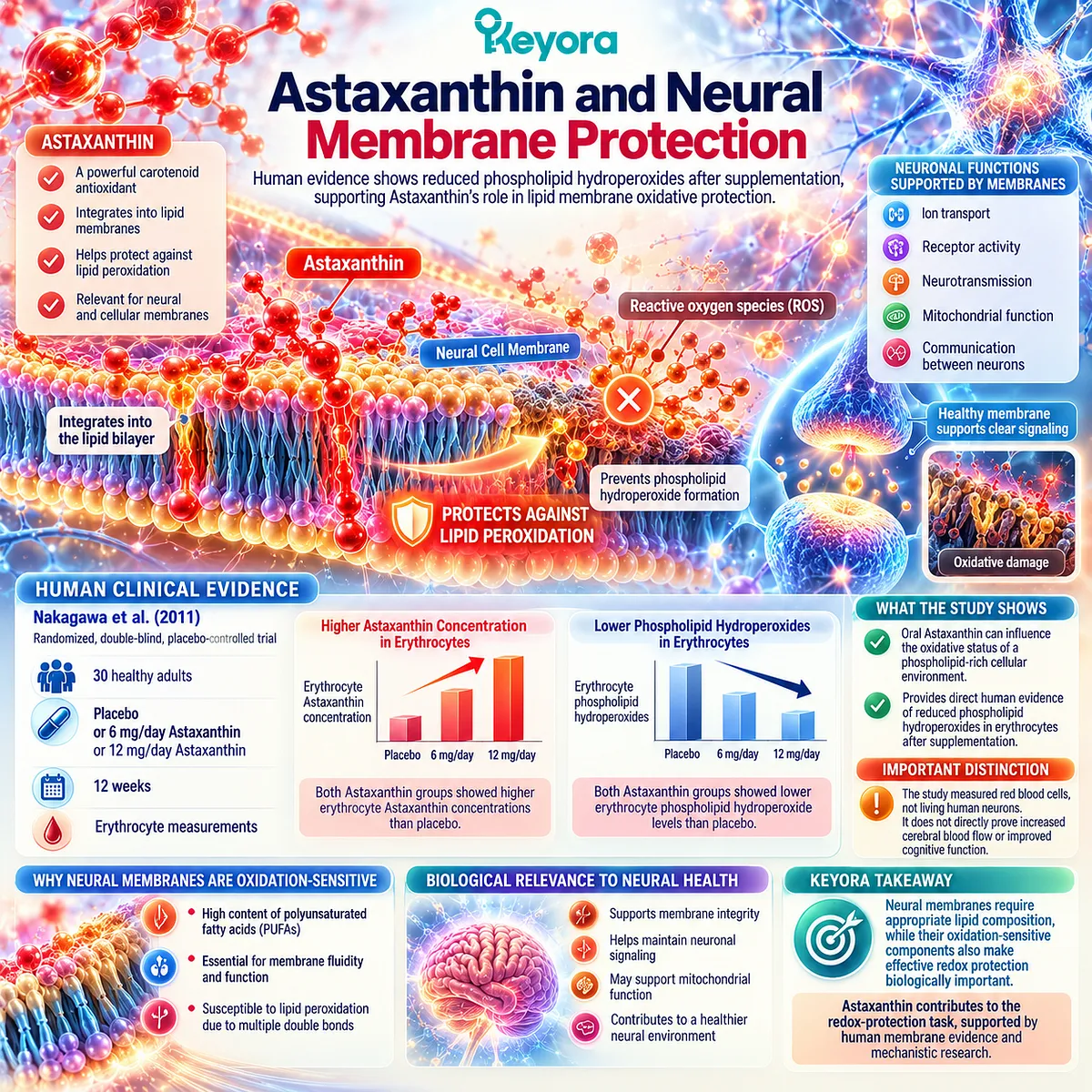
In a 2011 randomized, double-blind, placebo-controlled trial, Nakagawa and colleagues studied 30 healthy adults receiving placebo, 6 mg Astaxanthin, or 12 mg Astaxanthin daily for 12 weeks.
Both Astaxanthin groups showed higher erythrocyte Astaxanthin concentrations and lower erythrocyte phospholipid hydroperoxide levels than placebo.
This provides direct human evidence that orally consumed Astaxanthin can influence the oxidative status of a phospholipid-rich cellular environment.
The distinction is that researchers measured red blood cells, not living human neurons.
Astaxanthin’s relevance to neural membranes is therefore supported by combining human membrane-redox evidence with experimental research on lipid bilayers and cellular oxidative protection.
This question develops the lipid-protection architecture presented in Keyora Astaxanthin EP-5: The Neural Fortress: A Mechanistic Analysis of Astaxanthin in Lipidomics Re-engineering and Neural Oxidative Debt.
The central nutritional principle is straightforward:
Neural membranes require appropriate lipid composition, while their oxidation-sensitive components also make effective redox protection biologically important.
Astaxanthin contributes to the second task.
Why Brain Cell Membranes Need Lipid Protection
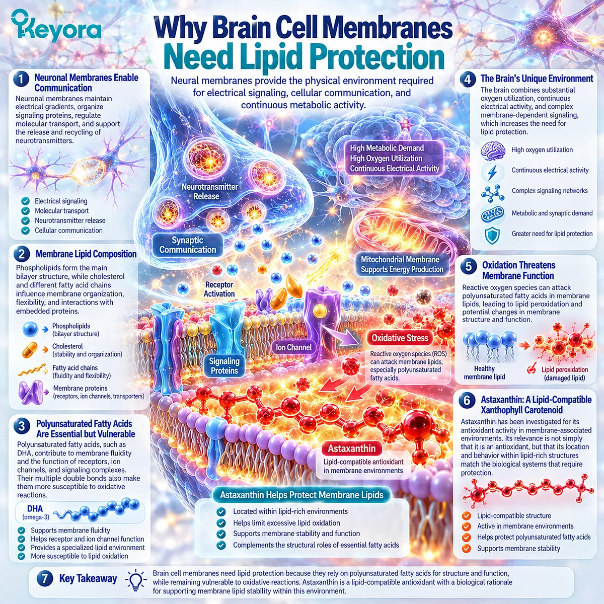
Neural membranes provide the physical environment required for electrical signaling, cellular communication, and continuous metabolic activity
Every thought depends on communication between specialized neural cells.
For that communication to occur, neuronal membranes must maintain electrical gradients, organize signaling proteins, regulate molecular transport, and support the repeated release and recycling of neurotransmitters.
These functions depend partly on membrane lipid composition.
Phospholipids form the main bilayer structure, while cholesterol and different fatty acid chains influence membrane organization, flexibility, and interactions with embedded proteins.
Polyunsaturated fatty acids are especially relevant because their multiple double bonds influence the physical behavior of lipid-rich biological structures.
DHA, for example, is a major long-chain omega-3 fatty acid in neural membrane biology. Its structural properties contribute to the environment in which receptors, ion channels, and signaling complexes operate.
Yet the same unsaturation that gives polyunsaturated fatty acids their distinctive physical properties also makes them more susceptible to oxidative reactions.
This creates a biological requirement that is easy to overlook.
Supplying appropriate lipids is one nutritional task. Maintaining a cellular environment capable of limiting excessive lipid oxidation is another.
The brain is particularly relevant to this relationship because it combines substantial oxygen utilization with continuous electrical activity and complex membrane-dependent signaling.
Mitochondrial membranes and synaptic membranes must also remain functional under changing metabolic demands.
Astaxanthin enters this discussion because it is a lipid-compatible xanthophyll carotenoid whose antioxidant activity has been investigated in membrane-associated environments.
Its relevance is therefore not simply that it belongs to the broad category of antioxidants.
It is that the location and behavior of an antioxidant matter when the biological structures requiring protection are themselves lipid-rich.
What Oxidative Stress Does to Neural Membrane Lipids
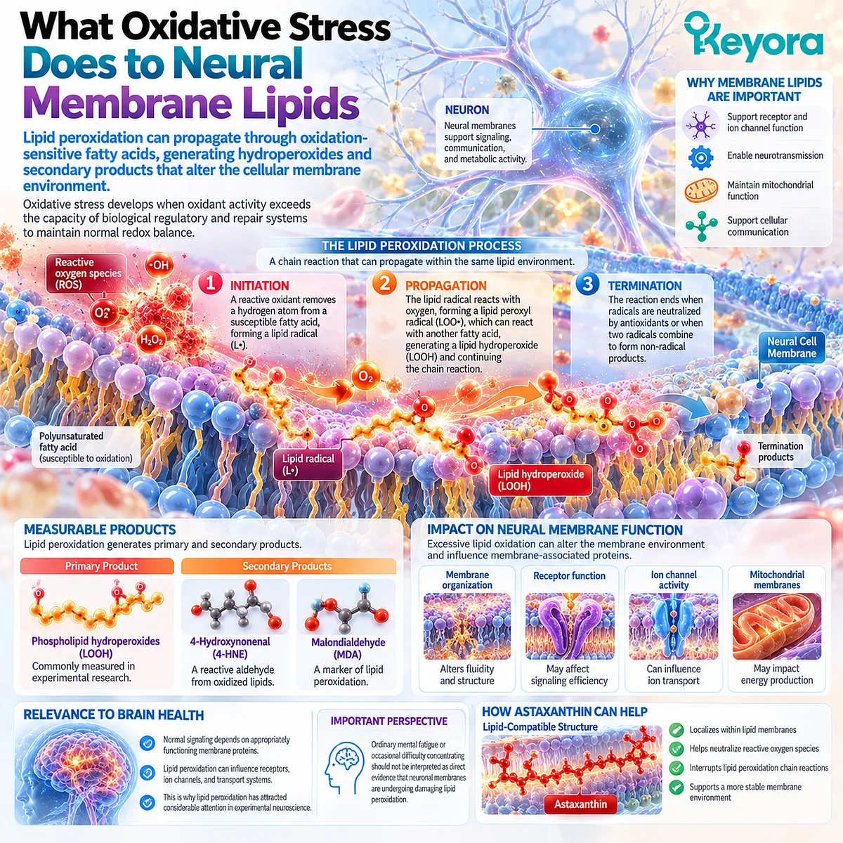
Lipid peroxidation can propagate through oxidation-sensitive fatty acids, generating hydroperoxides and secondary products that alter the cellular membrane environment
Oxidative stress develops when oxidant activity exceeds the capacity of biological regulatory and repair systems to maintain normal redox balance.
Within a lipid-rich membrane, one particularly important process is lipid peroxidation.
It begins when a sufficiently reactive oxidant initiates a reaction involving a susceptible fatty acid chain.
The resulting lipid radical can react with molecular oxygen, forming a lipid peroxyl radical. This intermediate may then react with another fatty acid, generating a lipid hydroperoxide and potentially propagating the reaction.
The process is commonly described through three stages:
Initiation – Propagation – Termination
Unlike a single isolated oxidation event, a propagating lipid reaction can involve neighboring molecules within the same lipid environment.
Phospholipid hydroperoxides are among the products that researchers can measure when investigating this process.
Further decomposition of oxidized lipids may produce reactive secondary compounds, including 4-hydroxynonenal (4-HNE) and malondialdehyde (MDA).
These molecules are biologically relevant because excessive lipid oxidation can influence membrane organization and interactions involving membrane-associated proteins.
For neural cells, those interactions matter because normal signaling depends on appropriately functioning receptors, ion channels, transport systems, and mitochondrial membranes.
This is why lipid peroxidation has attracted considerable attention in experimental neuroscience.
However, ordinary mental fatigue or occasional difficulty concentrating should not be interpreted as direct evidence that someone’s neuronal membranes are undergoing damaging lipid peroxidation.
The scientific question is more useful when focused on the mechanism itself:
Can a lipid-compatible nutritional compound help influence an oxidation-sensitive membrane environment?
Astaxanthin has both experimental and human biomarker evidence relevant to that question.
What Phospholipid Hydroperoxides Actually Measure
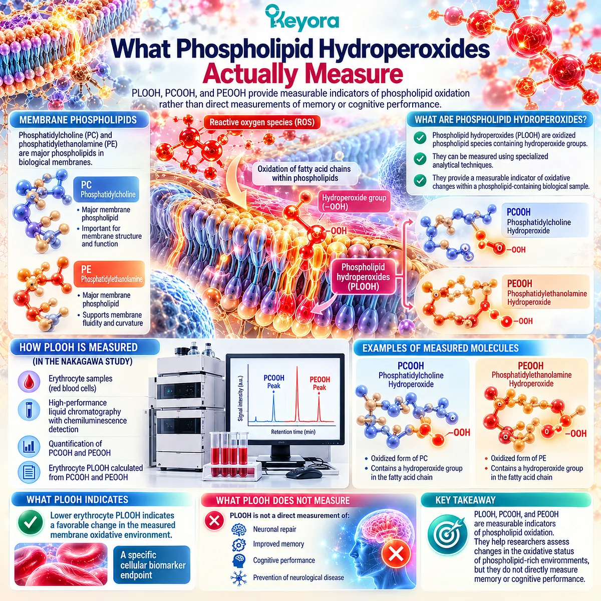
PLOOH, PCOOH, and PEOOH provide measurable indicators of phospholipid oxidation rather than direct measurements of memory or cognitive performance
Phospholipid hydroperoxides are commonly abbreviated as PLOOH.
They represent oxidized phospholipid species containing hydroperoxide groups and can be measured using specialized analytical techniques.
Two important examples are:
-
PCOOH – Phosphatidylcholine Hydroperoxide
-
PEOOH – Phosphatidylethanolamine Hydroperoxide
Phosphatidylcholine and phosphatidylethanolamine are major classes of biological membrane phospholipids.
When fatty acid components within these molecules undergo oxidation, corresponding phospholipid hydroperoxides may form.
Measuring these products provides researchers with information about oxidative changes occurring within a phospholipid-containing biological sample.
This approach is particularly useful because it moves beyond the generalized statement that a substance has antioxidant activity in a laboratory chemical assay.
Instead, researchers can investigate whether oral supplementation is associated with measurable changes in oxidized lipid species within human cells.
In the Nakagawa trial, erythrocyte PLOOH was calculated using measurements of PCOOH and PEOOH. Researchers used high-performance liquid chromatography with chemiluminescence detection to quantify these oxidation products.
This provided a specific cellular biomarker endpoint.
The distinction between biomarker and clinical outcome remains important:
Lower erythrocyte PLOOH indicates a favorable change in the measured membrane oxidative environment.
It is not, by itself, a direct measurement of neuronal repair, improved memory, or prevention of neurological disease.
What Human Astaxanthin Research Actually Shows
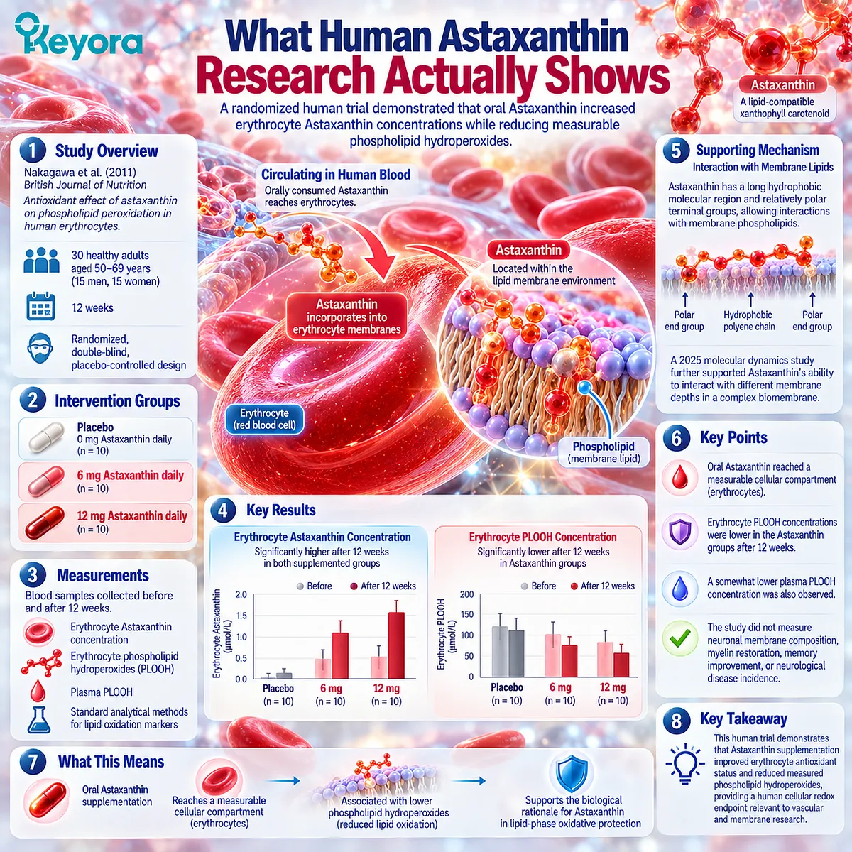
A randomized human trial demonstrated that oral Astaxanthin increased erythrocyte Astaxanthin concentrations while reducing measurable phospholipid hydroperoxides
The principal human evidence for this question comes from Nakagawa and colleagues, published in the British Journal of Nutrition in 2011.
The study was titled Antioxidant effect of astaxanthin on phospholipid peroxidation in human erythrocytes.
Researchers recruited 30 healthy adults aged 50 – 69 years, including 15 men and 15 women.
Participants were randomly assigned to one of three interventions:
-
Placebo – 0 mg Astaxanthin
-
6 mg Astaxanthin daily
-
12 mg Astaxanthin daily
The intervention continued for 12 weeks under a double-blind, placebo-controlled design.
Blood samples were collected before supplementation and at the end of the study. Researchers measured erythrocyte Astaxanthin concentrations alongside phospholipid oxidation products.
The results showed that erythrocyte Astaxanthin concentrations were significantly higher in both supplemented groups than in placebo.
More importantly, erythrocyte PLOOH concentrations were lower in the Astaxanthin groups after supplementation.
A somewhat lower plasma PLOOH concentration was also observed, although the erythrocyte findings provided the more direct cellular membrane endpoint.
These results establish two connected observations.
-
First, orally consumed Astaxanthin reached a measurable cellular compartment.
-
Second, its presence was associated with a reduction in specific phospholipid oxidation products within that compartment.
This is scientifically meaningful because it connects oral nutritional intervention with a human cellular redox outcome, rather than relying exclusively on chemical antioxidant comparisons.
The original researchers discussed the potential relevance of their findings to conditions associated with elevated oxidative stress. Nevertheless, the study did not measure neuronal membrane composition, myelin restoration, memory improvement, or neurological disease incidence.
Its strongest conclusion is therefore:
Astaxanthin supplementation improved erythrocyte antioxidant status and reduced measured phospholipid hydroperoxides in this human intervention.
Experimental lipid-bilayer research provides a complementary explanation.
Astaxanthin possesses a long hydrophobic molecular region and relatively polar terminal groups. This combination enables interactions with membrane phospholipids, although its exact location and orientation depend on the experimental membrane environment.
A 2025 molecular dynamics study further investigated Astaxanthin’s location and behavior in a complex biomembrane, supporting its ability to interact with different membrane depths.
Together, these findings support the biological rationale for Astaxanthin in lipid-phase oxidative protection.
Where Keyora Asta 16MG and ALA Fit
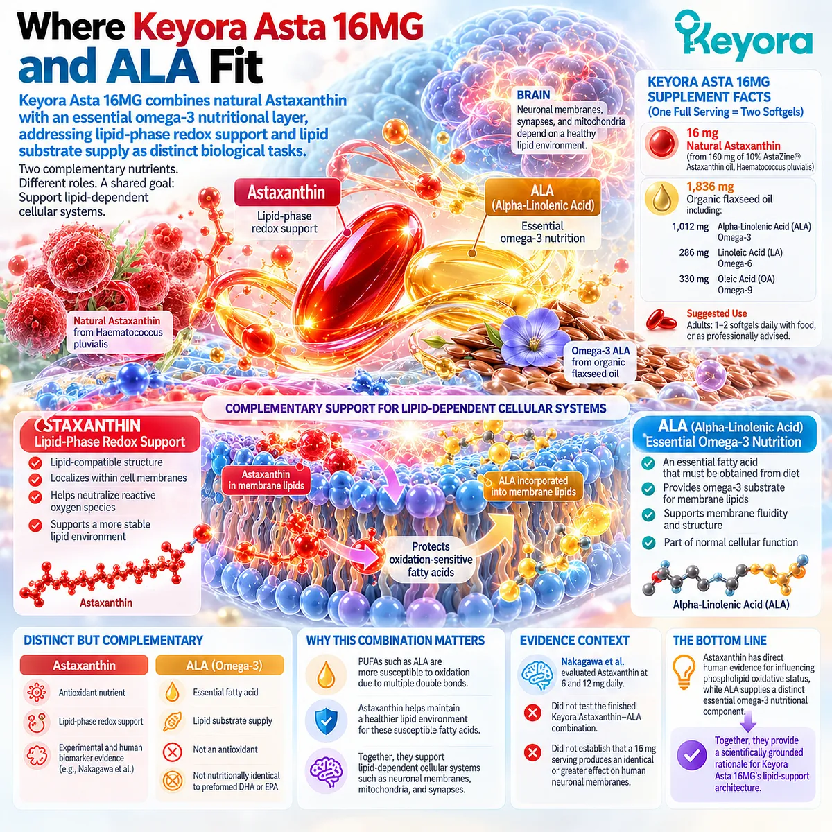
Keyora Asta 16MG combines natural Astaxanthin with an essential omega-3 nutritional layer, addressing lipid-phase redox support and lipid substrate supply as distinct biological tasks
The current Keyora Asta 16MG Supplement Facts define one full serving as two softgels.
That serving provides:
16 mg Natural Astaxanthin, supplied by 160 mg of 10% AstaZine® Astaxanthin oil derived from Haematococcus pluvialis.
The formula also contains 1,836 mg organic flaxseed oil, including:
-
1,012 mg Alpha-Linolenic Acid (ALA), Omega-3
-
286 mg Linoleic Acid (LA), Omega-6
-
330 mg Oleic Acid (OA), Omega-9
The suggested adult use is one to two softgels daily with food, or as professionally advised.
This composition introduces an important nutritional distinction.
-
Astaxanthin is the lipid-compatible redox-support nutrient.
-
ALA is an essential omega-3 polyunsaturated fatty acid that the human body must obtain through dietary intake.
-
ALA is not simply an alternative name for an antioxidant, nor is it nutritionally identical to preformed DHA or EPA.
Its principal role within this discussion is to provide a separate essential fatty acid layer, while Astaxanthin contributes antioxidant properties relevant to lipid-rich biological environments.
The Keyora Lipid-Phase Oxidative Protection framework can therefore be expressed as:
-
Essential Lipid Nutrition – ALA
-
Lipid-Phase Redox Support – Astaxanthin
↓
-
Complementary Support for Lipid-Dependent Cellular Systems
This relationship is especially relevant when discussing polyunsaturated fatty acids, because their multiple double bonds make the surrounding lipid environment susceptible to oxidation.
It also explains why ALA should not be reduced to an inactive carrier oil within the Keyora formula.
However, the Nakagawa trial evaluated Astaxanthin at 6 and 12 mg daily. It did not test the finished Keyora Astaxanthin-ALA combination or demonstrate that a 16 mg serving produces an identical or greater effect on human neuronal membranes.
The evidence-aligned interpretation remains clear.
Astaxanthin has direct human evidence for influencing phospholipid oxidative status, while ALA supplies a distinct essential omega-3 nutritional component.
Together, these complementary nutritional tasks provide the biological rationale for Keyora Asta 16MG’s lipid-support architecture.
The relevant goal is not to claim that an antioxidant can permanently shield every brain cell from oxidation.
It is to recognize that neural function depends on lipid-rich cellular structures, and that maintaining the biological environment surrounding those structures is a meaningful target for nutritional research.
This article is for educational and informational purposes only. It does not provide medical advice, diagnosis, treatment, cure, prevention, disease outcome claims, hormone restoration claims, fertility outcome claims, or formula-specific clinical efficacy claims.