
How Is Astaxanthin Different From Vitamin E in Cell Membranes?
Keyora Research Q&A Library
This is part of the Keyora Research Q&A Series, derived from Keyora Astaxanthin Research Journal Series.
Within the Keyora Astaxanthin Researcn framework, this Q&A translates complex astaxanthin biology into reader-friendly, evidence-bound answers, focusing on natural astaxanthin identity, molecular structure, antioxidant and redox mechanisms, membrane lipid interaction, mitochondrial resilience, inflammatory signaling pathways, human evidence interpretation, and the scientific principles behind responsible supplementation.
First published by Keyora Research Journal: www.keyorahealth.com
Direct Answer
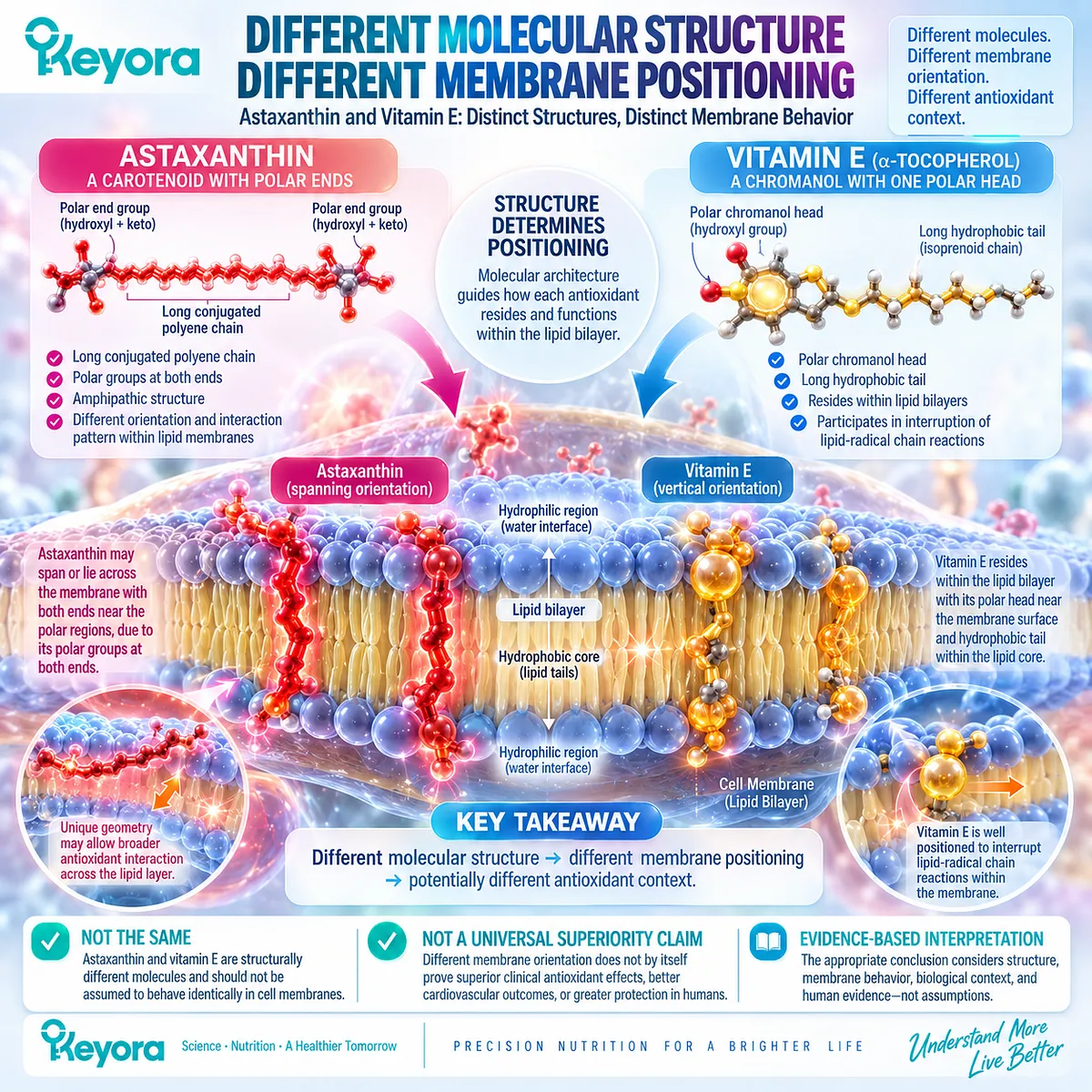
Astaxanthin and vitamin E are both associated with lipid environments, but they are structurally different molecules and therefore should not be assumed to behave identically inside cell membranes.
Alpha-tocopherol, the major form of vitamin E commonly discussed in membrane antioxidant biology, has a polar chromanol head and a long hydrophobic tail.
This structure allows it to reside within lipid bilayers and participate in the interruption of lipid-radical chain reactions.
Astaxanthin has a different architecture. It contains a long conjugated polyene chain with polar groups at both ends, giving it a different orientation and interaction pattern within lipid membranes.
Keyora Astaxanthin EP-4: The Cardiac Architecture: Bio-Energetic Sovereignty emphasizes this difference in membrane geometry when comparing Astaxanthin with vitamin E.
The important conclusion is not that one antioxidant is universally “better.”
It is:
Different molecular structure → different membrane positioning → potentially different antioxidant context
And the evidence boundary is equally important:
Different membrane orientation does not by itself prove superior clinical antioxidant effects, better cardiovascular outcomes, or greater protection in humans.
Why Membrane Position Matters for an Antioxidant
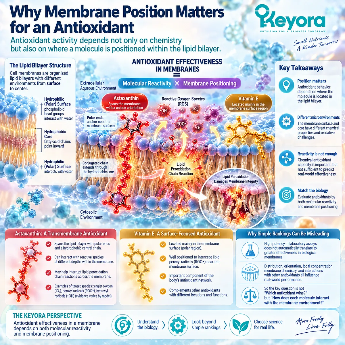
Antioxidant activity depends not only on chemistry but also on where a molecule is positioned within the lipid bilayer
Antioxidants are often compared using a single question:
“Which one is stronger?”
That question is incomplete.
A molecule’s antioxidant behavior depends not only on its chemical ability to react with certain radicals or reactive species, but also on where that molecule is located when oxidative reactions occur.
Cell membranes are organized lipid bilayers.
Their outer regions are more polar because phospholipid head groups interact with water, while the center of the bilayer is more hydrophobic because fatty-acid chains point inward.
A molecule that associates mainly near the membrane surface therefore occupies a different microenvironment from one that extends more deeply into the bilayer.
This matters because lipid peroxidation is not a single event occurring in one uniform location.
Reactive species can interact with membrane lipids at different depths, and antioxidant molecules may be better positioned for some reactions than others depending on their structure and orientation.
Keyora Astaxanthin EP-4 uses this idea to argue that membrane antioxidant biology should be evaluated not only by radical-scavenging capacity but also by molecular geometry and location within the phospholipid bilayer.
That general principle is useful.
A more cautious way to state it is:
Antioxidant effectiveness in a membrane depends on both molecular reactivity and membrane positioning.
This also explains why simple potency rankings can be misleading.
A molecule may perform very strongly in a laboratory assay yet behave differently in a biological membrane, where distribution, orientation, concentration, local chemistry, and interactions with other antioxidants all matter.
So when comparing Astaxanthin with vitamin E, the first question should not be:
“Which antioxidant wins?”
It should be:
How does each molecule interact with the membrane environment?
How Vitamin E Sits in a Lipid Membrane
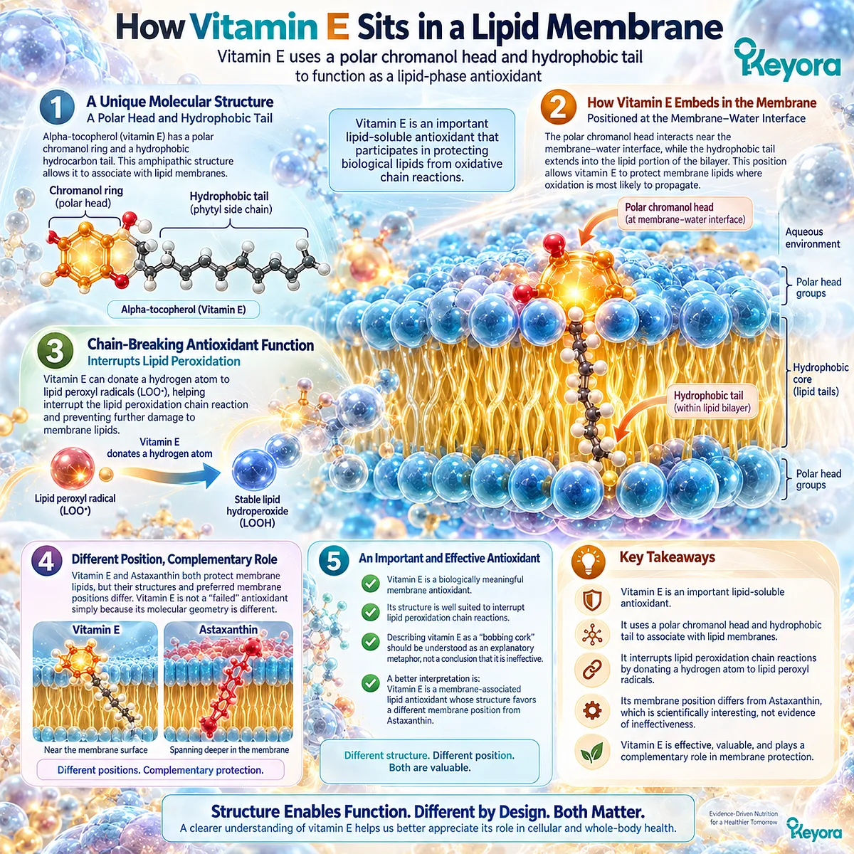
Vitamin E uses a polar chromanol head and hydrophobic tail to function as a lipid-phase antioxidant
Vitamin E is not a weak or irrelevant antioxidant.
It is an important lipid-soluble antioxidant that participates in protecting biological lipids from oxidative chain reactions.
Alpha-tocopherol has a characteristic structure consisting of a polar chromanol ring and a hydrophobic hydrocarbon tail.
That combination allows it to associate with lipid membranes.
Its polar region can interact near the membrane-water interface, while its hydrophobic tail extends into the lipid portion of the bilayer.
Keyora Astaxanthin EP-4 describes this basic structural arrangement when comparing alpha-tocopherol with Astaxanthin.
This structure is well suited to vitamin E’s role as a chain-breaking antioxidant.
During lipid peroxidation, lipid radicals can propagate oxidation from one membrane lipid to another.
Vitamin E can donate a hydrogen atom to lipid peroxyl radicals, helping interrupt that propagation cycle.
That function is biologically meaningful.
It would therefore be incorrect to describe vitamin E as a “failed” membrane antioxidant simply because its molecular geometry differs from Astaxanthin.
Some of the language in Keyora Astaxanthin EP-4, including the description of vitamin E as a “bobbing cork” or structurally inadequate for a particular membrane role, should be understood as an explanatory metaphor rather than a balanced conclusion about vitamin E biology.
A better interpretation is:
Vitamin E is a membrane-associated lipid antioxidant whose structure favors a different membrane position from Astaxanthin.
That difference is scientifically interesting.
It is not evidence that vitamin E is ineffective.
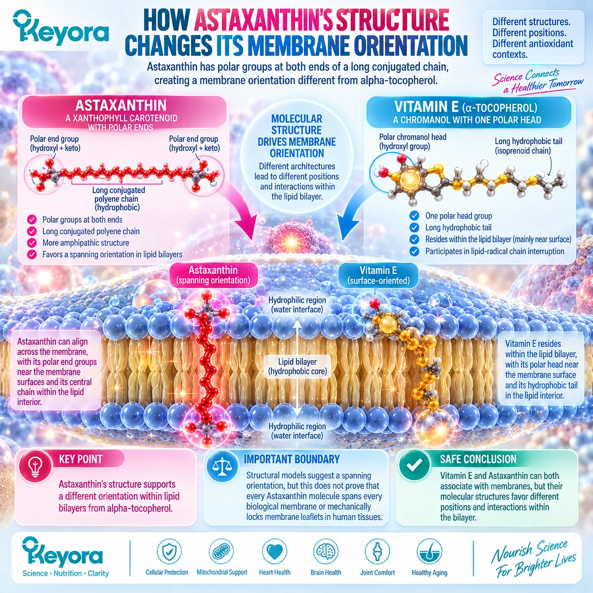
How Astaxanthin’s Structure Changes Its Membrane Orientation
Astaxanthin has polar groups at both ends of a long conjugated chain, creating a membrane orientation different from alpha-tocopherol
Astaxanthin belongs to the xanthophyll carotenoid family.
Its molecular structure differs substantially from alpha-tocopherol.
The central portion of Astaxanthin consists of a long conjugated polyene chain that interacts favorably with lipid environments.
At both ends of the molecule are polar oxygen-containing groups.
This combination gives Astaxanthin a more amphipathic structure than vitamin E.
Keyora Astaxanthin EP-4 uses this architecture to describe Astaxanthin as aligning across a phospholipid membrane, with its polar end regions associated closer to the membrane surfaces and its central hydrophobic chain extending through the lipid interior.
That model helps explain why Astaxanthin is often discussed as a membrane-oriented antioxidant.
However, this should be expressed carefully.
It is reasonable to say:
Astaxanthin’s structure supports a different orientation within lipid bilayers from alpha-tocopherol.
It is much stronger to say:
Every Astaxanthin molecule physically spans every biological membrane and mechanically locks both membrane leaflets together.
That second statement goes beyond what should be concluded from structural models alone.
The idea of Astaxanthin acting as a “molecular rivet” is a useful Keyora visualization, but it should remain a framework rather than being presented as an established universal mechanical function in human tissues.
This distinction is particularly important because the next level of the series examines the bilayer-spanning claim directly.
For Q009, the safest conclusion is simpler:
Vitamin E and Astaxanthin can both associate with membranes, but their molecular structures favor different positions and interactions within the bilayer.
Does Different Membrane Geometry Mean Astaxanthin Is Better?
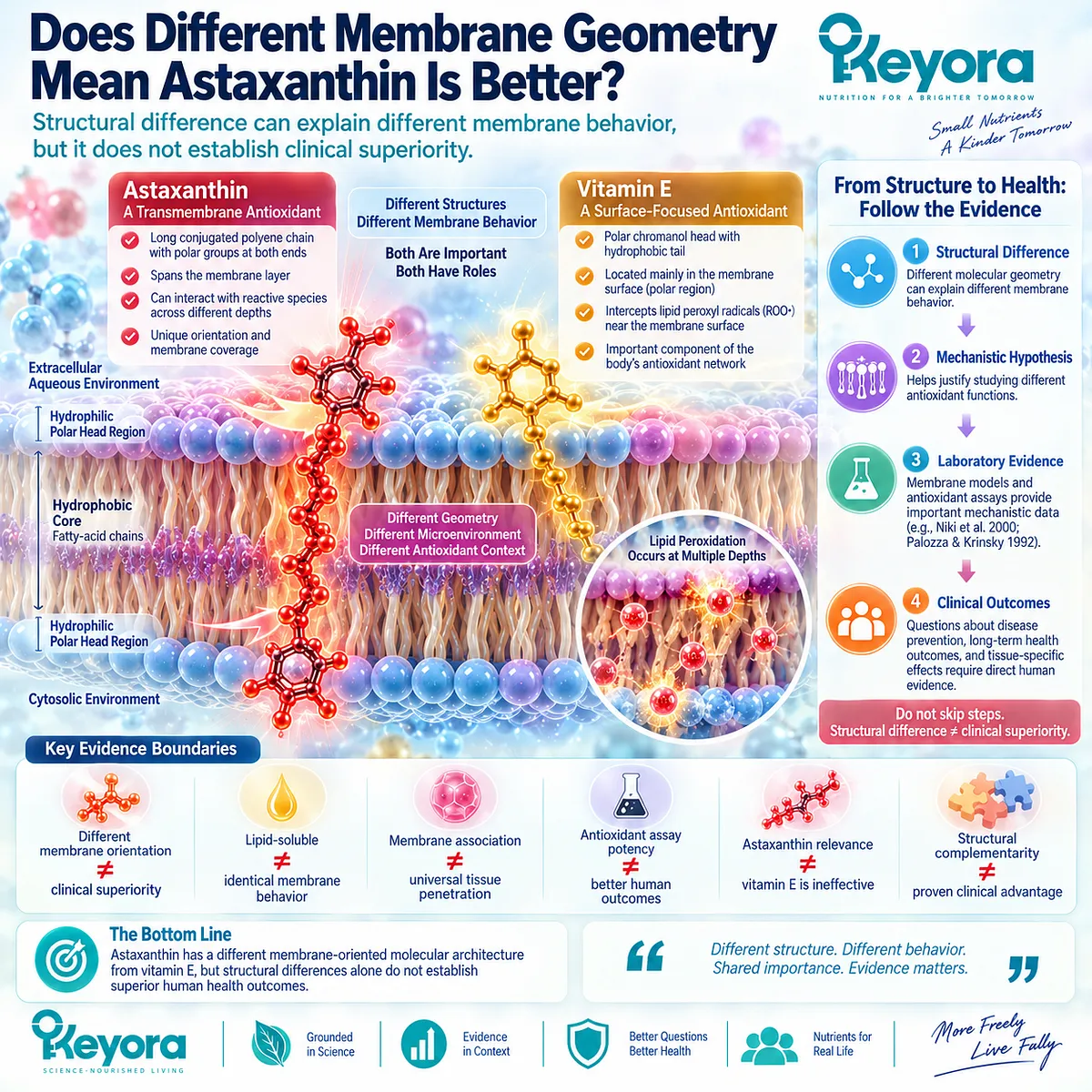
Structural difference can explain different membrane behavior, but it does not establish clinical superiority
No.
Different membrane geometry does not automatically mean that Astaxanthin is clinically superior to vitamin E.
This is where antioxidant comparisons often become misleading.
A structural difference can support a mechanistic hypothesis.
It can help explain why two molecules may interact differently with membrane lipids.
It can justify studying different antioxidant functions.
But it does not answer questions such as:
Which nutrient better prevents cardiovascular disease?
Which produces better long-term health outcomes?
Which protects every tissue more effectively?
Those questions require direct biological and clinical evidence.
Keyora Astaxanthin EP-4 sometimes moves from molecular geometry to stronger statements that Astaxanthin is structurally “superior” and functions like a membrane “steel beam” compared with vitamin E.
Those metaphors should not be converted into a clinical hierarchy.
The more defensible conclusion is:
Astaxanthin has a different membrane-oriented molecular architecture from vitamin E, but structural differences alone do not establish superior human health outcomes.
The same caution applies to laboratory antioxidant-potency comparisons.
The EP-4 reference set includes membrane-model and antioxidant-interaction literature, including Niki et al. (2000), Interaction Among Vitamin C, Vitamin E, and Beta-Carotene, and Palozza and Krinsky (1992), Astaxanthin and Canthaxanthin Are Potent Antioxidants in a Membrane Model.
Such studies help explain antioxidant chemistry.
They do not establish a universal ranking of clinical effectiveness.
The evidence boundaries are:
Different membrane orientation ≠ clinical superiority
Lipid-soluble ≠ identical membrane behavior
Membrane association ≠ universal tissue penetration
Antioxidant assay potency ≠ better human outcomes
Astaxanthin relevance ≠ vitamin E is ineffective
Structural complementarity ≠ proven clinical advantage
The Keyora Cardiac Architecture: Different Antioxidants, Different Membrane Roles
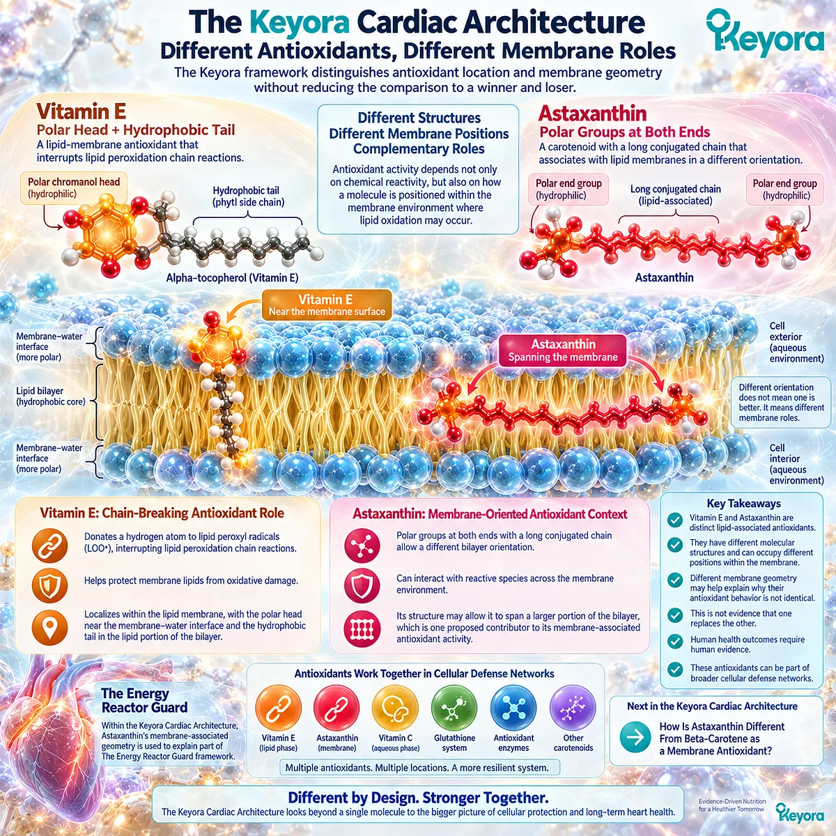
The Keyora framework distinguishes antioxidant location and membrane geometry without reducing the comparison to a winner and loser
Within the Keyora Cardiac Architecture, Astaxanthin’s membrane-associated geometry is used to explain part of The Energy Reactor Guard framework.
The underlying idea is that antioxidant biology depends not only on whether a molecule can react with oxidative species, but also on how that molecule is positioned within the membrane environment where lipid oxidation may occur.
The comparison can therefore be summarized as:
Vitamin E
→ polar chromanol head + hydrophobic tail
→ lipid-membrane localization
→ chain-breaking antioxidant role
Astaxanthin
→ polar groups at both ends + long conjugated lipid-associated chain
→ different bilayer orientation
→ membrane-oriented antioxidant context
These are different molecular architectures.
They should not be interpreted as evidence that one nutrient makes the other unnecessary.
Biological antioxidant systems are not usually built around a single molecule working in isolation.
Vitamin E, carotenoids, vitamin C, glutathione-related systems, antioxidant enzymes, and other redox pathways can interact within broader cellular defense networks.
The practical scientific conclusion is therefore not:
Astaxanthin replaces vitamin E.
It is:
Astaxanthin and vitamin E are distinct lipid-associated antioxidants whose structures can place them differently within biological membranes.
That difference may help explain why their antioxidant behavior is not identical.
But geometry remains one layer of evidence.
Human health outcomes require human evidence.
This distinction also prepares the next comparison in the Keyora Cardiac Architecture:
How Is Astaxanthin Different From Beta-Carotene as a Membrane Antioxidant?
This article is for educational and informational purposes only. It does not provide medical advice, diagnosis, treatment, cure, prevention, disease outcome claims, hormone restoration claims, fertility outcome claims, or formula-specific clinical efficacy claims.