
Why Is Astaxanthin Studied in So Many Tissues?
Keyora Research Q&A Library
This is part of the Keyora Research Q&A Series, derived from Keyora Astaxanthin Research Journal Series.
Within the Keyora Astaxanthin Researcn framework, this Q&A translates complex astaxanthin biology into reader-friendly, evidence-bound answers, focusing on natural astaxanthin identity, molecular structure, antioxidant and redox mechanisms, membrane lipid interaction, mitochondrial resilience, inflammatory signaling pathways, human evidence interpretation, and the scientific principles behind responsible supplementation.
First published by Keyora Research Journal: www.keyorahealth.com
Direct Answer
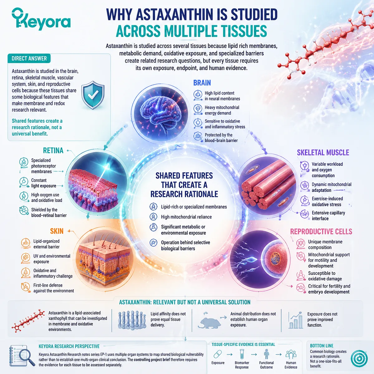
Astaxanthin is studied in the brain, retina, skeletal muscle, vascular system, skin, and reproductive cells because these tissues share some biological features that make membrane and redox research relevant.
They may contain specialized lipid structures, depend heavily on mitochondrial metabolism, encounter substantial metabolic or environmental exposure, or operate behind selective biological barriers.
Astaxanthin is studied across several tissues because lipid rich membranes, metabolic demand, oxidative exposure, and specialized barriers create related research questions, but every tissue requires its own exposure, endpoint, and human evidence.
Shared features create a research rationale, not a universal benefit.
The retina processes light and has highly specialized photoreceptor membranes.
-
Skeletal muscle changes its oxygen consumption and mitochondrial activity with workload.
-
Skin contains a lipid organized external barrier.
-
Sperm and oocytes have distinct membrane, mitochondrial, and developmental requirements.
These tissues cannot be treated as interchangeable versions of one redox system.
Astaxanthin is relevant because it is a lipid associated xanthophyll that can be investigated in membrane and oxidative environments.
However, lipid affinity does not prove equal tissue delivery.
Animal distribution does not establish human organ exposure, and exposure does not prove improved function.
Keyora Astaxanthin Research notes series EP-1 uses multiple organ systems to map shared biological vulnerability rather than to establish one multi-organ clinical conclusion.
The controlling project brief therefore requires the evidence for each tissue to be assessed separately.
Shared Features Create Research Interest
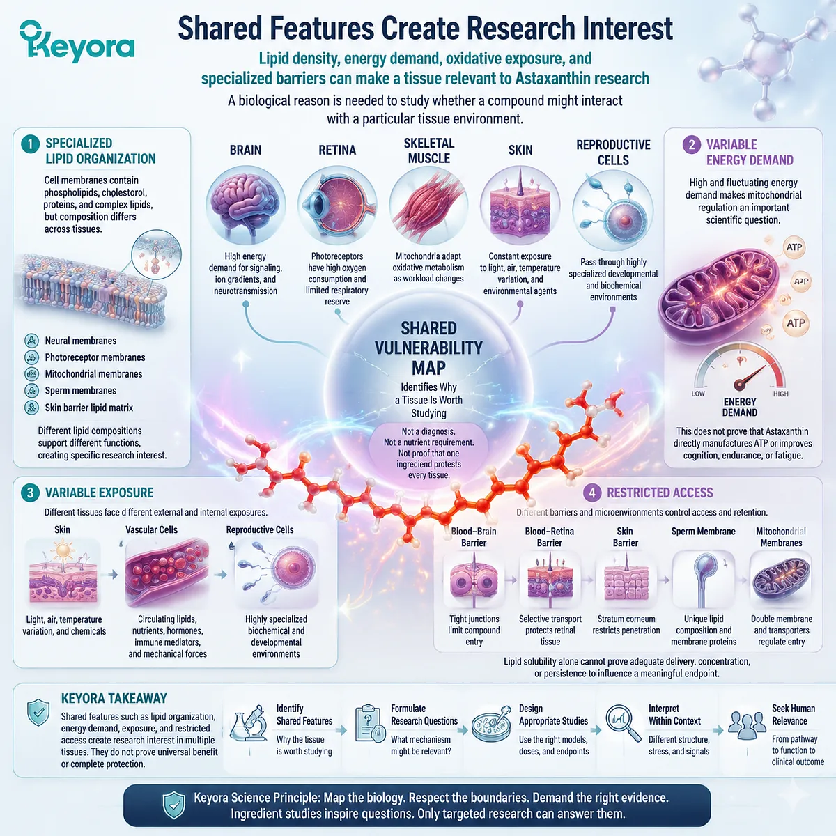
Lipid density, energy demand, oxidative exposure, and specialized barriers can make a tissue relevant to Astaxanthin research
A nutrient does not become scientifically relevant to several tissues merely because those tissues belong to the same body.
Researchers usually need a biological reason to ask whether a compound might interact with a particular tissue environment.
One reason is specialized lipid organization.
Cell membranes contain phospholipids, cholesterol, proteins, and other complex lipids, but their composition is not identical across tissues.
Neural membranes, photoreceptor membranes, mitochondrial membranes, sperm membranes, and the lipid matrix of the skin barrier perform different tasks.
The retina offers a clear example. Photoreceptor outer segments contain specialized membranes that support the conversion of light into neural signals.
Experimental measurements of intact retina have demonstrated substantial oxygen consumption and limited respiratory reserve in photoreceptors, creating interest in mitochondrial and redox regulation within this tissue.
This does not mean that every antioxidant improves vision. It explains why retinal metabolism and membrane biology are frequently studied.
A second reason is variable energy demand.
Brain signaling requires considerable energy for ion gradients, action potentials, neurotransmission, and cellular maintenance.
Skeletal muscle mitochondria must adjust oxidative metabolism as workload changes.
These energetic demands make mitochondrial regulation an important scientific question, but they do not prove that Astaxanthin directly manufactures ATP or improves cognition, endurance, or fatigue.
A third reason is exposure.
-
Skin faces light, air, temperature variation, and environmental substances.
-
Vascular cells encounter circulating lipids, nutrients, hormones, immune mediators, and mechanical forces.
-
Reproductive cells pass through highly specialized developmental and biochemical environments.
A fourth reason is restricted access.
The brain, retina, skin, reproductive tract, and individual cell organelles have different transport conditions and local microenvironments.
Lipid solubility alone cannot prove that Astaxanthin crosses each barrier, reaches an adequate concentration, or remains available long enough to influence a meaningful endpoint.
These shared features form what Keyora can describe as a Shared Vulnerability Map.
The map identifies why a tissue is worth studying. It does not diagnose vulnerability, prove a nutrient requirement, or establish that one ingredient protects every listed tissue.
Tissue Specialization Changes the Scientific Question
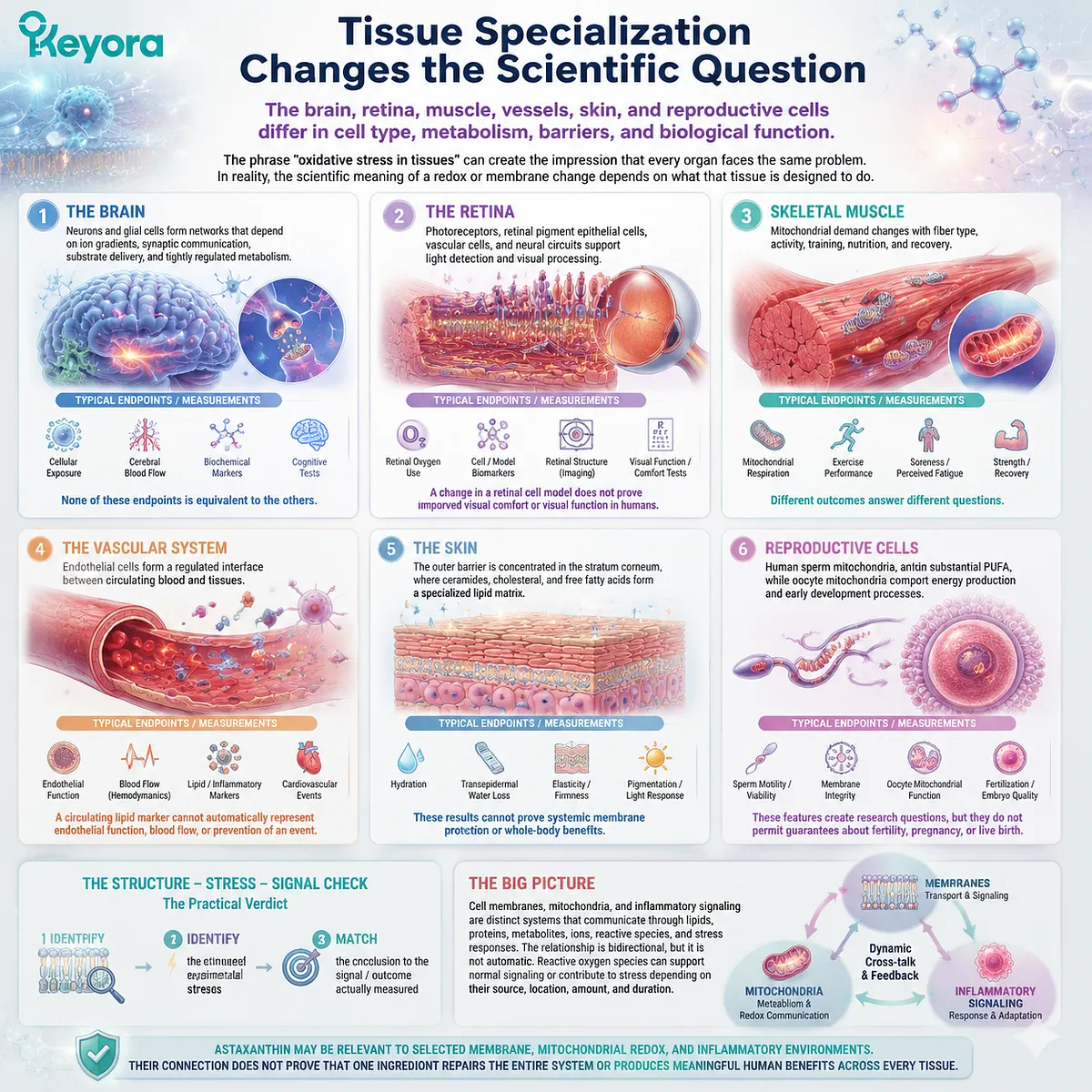
The brain, retina, muscle, vessels, skin, and reproductive cells differ in cell type, metabolism, barriers, and biological function
The phrase “oxidative stress in tissues” can create the impression that every organ faces the same problem. In reality, the scientific meaning of a redox or membrane change depends on what that tissue is designed to do.
In the brain, neurons and glial cells form networks that depend on ion gradients, synaptic communication, substrate delivery, and tightly regulated metabolism.
A brain study might examine cellular exposure, blood flow, a biochemical marker, or a cognitive test.
None of these endpoints is equivalent to the others.
In the retina, photoreceptors, retinal pigment epithelial cells, vascular cells, and neural circuits support light detection and visual processing.
Retinal oxygen use and specialized photoreceptor membranes make redox biology relevant, but a change in a retinal cell model does not prove improved visual comfort or visual function in humans.
In skeletal muscle, mitochondrial demand changes with fiber type, activity, training, nutrition, and recovery.
Mitochondrial respiration may be measured directly in muscle samples, while human studies may instead assess exercise performance, soreness, perceived fatigue, strength, or recovery. These outcomes answer different questions.
The vascular system introduces another context.
Endothelial cells form a regulated interface between circulating blood and tissues.
Red blood cells, vascular smooth muscle, plasma lipoproteins, and immune cells create additional targets and measurements.
A circulating lipid marker cannot automatically represent endothelial function, blood flow, or prevention of a cardiovascular event.
Skin differs again. Its outer barrier is concentrated in the stratum corneum, where ceramides, cholesterol, and free fatty acids form a specialized lipid matrix.
This structure is not equivalent to the phospholipid membrane of a neuron or muscle cell.
Skin studies may measure hydration, transepidermal water loss, elasticity, pigmentation, or responses to light exposure, but such results cannot prove systemic membrane protection.
Reproductive cells are also highly specialized.
Human sperm membranes contain substantial polyunsaturated fatty acids, while sperm motility is connected to mitochondrial and membrane function.
Oocyte mitochondria support energy production and other processes required during maturation and early development. These features create research questions, but they do not permit guarantees about fertility, pregnancy, or live birth.
The same broad mechanism can therefore lead researchers toward very different measurements. This article does not determine whether Astaxanthin improves those endpoints. It establishes why the endpoints must remain tissue specific.
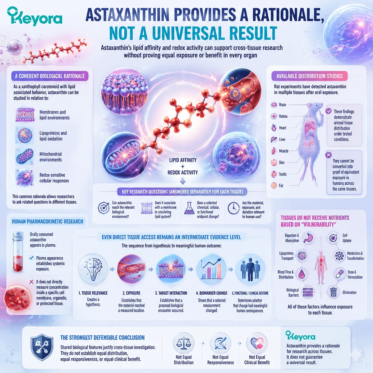
Astaxanthin Provides a Rationale, Not a Universal Result
Astaxanthin’s lipid affinity and redox activity can support cross-tissue research without proving equal exposure or benefit in every organ
Astaxanthin’s identity provides a coherent reason for its appearance across different research fields.
As a xanthophyll carotenoid with lipid associated behavior, it can be studied in relation to membranes, lipoproteins, lipid oxidation, mitochondrial environments, and selected redox sensitive responses.
That common rationale is scientifically useful. It allows researchers to ask related questions in different tissues:
-
Can Astaxanthin reach the relevant biological environment?
-
Does it associate with a membrane or circulating lipid system?
-
Does a selected chemical, cellular, or functional endpoint change?
-
Are the material, exposure, and duration relevant to human use?
These questions must be answered separately for each tissue.
Available distribution studies illustrate the problem.
Rat experiments have detected Astaxanthin in several tissues after oral exposure, with concentrations and isomer patterns differing among tissues.
These findings demonstrate animal tissue distribution under the tested formulations and doses. They cannot be converted into proof that ordinary human supplementation provides equivalent exposure to the brain, retina, skin, muscle, vascular system, or reproductive cells.
Human pharmacokinetic research can demonstrate that orally consumed Astaxanthin appears in plasma.
Plasma appearance establishes systemic exposure, but it does not directly measure concentration inside a specific cell membrane, organelle, or protected tissue.
Even direct tissue access would remain an intermediate evidence level. The sequence is:
Tissue relevance creates a hypothesis.
Exposure establishes that the material reached a measured location.
Target interaction establishes that a proposed biological encounter occurred.
A biomarker shows that a selected measurement changed.
A functional or clinical study determines whether that change had meaningful human consequences.
Astaxanthin should therefore not be presented as a compound that automatically seeks out every lipid rich or energy demanding tissue.
Tissues do not receive nutrients simply according to which one appears most vulnerable.
Digestion, transport, lipoproteins, metabolism, blood flow, barriers, cell uptake, dose, formulation, and elimination all influence exposure.
The strongest defensible conclusion is that shared biological features justify cross-tissue investigation. They do not establish equal distribution, equal responsiveness, or equal clinical benefit.
Use the Keyora Shared Feature – Tissue – Evidence Check
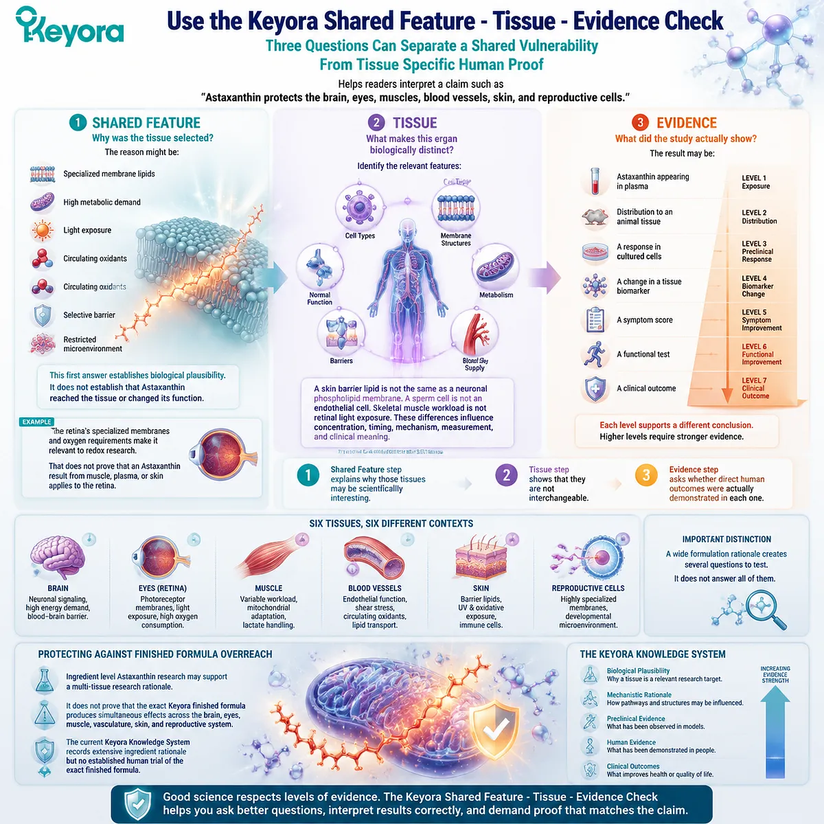
Three questions can separate a shared vulnerability from tissue specific human proof
The Keyora Shared Feature – Tissue – Evidence Check helps readers interpret a claim such as “Astaxanthin protects the brain, eyes, muscles, blood vessels, skin, and reproductive cells.”
Shared Feature: Why was the tissue selected?
The reason might be specialized membrane lipids, high metabolic demand, light exposure, circulating oxidants, a selective barrier, or a restricted microenvironment.
This first answer establishes biological plausibility. It does not establish that Astaxanthin reached the tissue or changed its function.
For example, the retina’s specialized membranes and oxygen requirements make it relevant to redox research. That does not prove that an Astaxanthin result from muscle, plasma, or skin applies to the retina.
Tissue: What makes this organ biologically distinct?
The reader should identify the relevant cell types, membrane structures, metabolism, blood supply, barriers, and normal function.
-
A skin barrier lipid is not the same as a neuronal phospholipid membrane.
-
A sperm cell is not an endothelial cell.
-
Skeletal muscle workload is not retinal light exposure.
These differences influence concentration, timing, mechanism, measurement, and clinical meaning.
Evidence: What did the study actually show?
The result may be:
-
Astaxanthin appearing in plasma
-
distribution to an animal tissue
-
a response in cultured cells
-
a change in a tissue biomarker
-
a symptom score
-
a functional test
-
a clinical outcome
Each level supports a different conclusion.
Consider the claim, “Astaxanthin is studied in six tissues, so it provides whole body protection.”
The Shared Feature step explains why those tissues may be scientifically interesting.
The Tissue step shows that they are not interchangeable.
The Evidence step asks whether direct human outcomes were actually demonstrated in each one.
This method also protects against finished formula overreach. Ingredient level Astaxanthin research may support a multi-tissue research rationale, but it does not prove that the exact Keyora finished formula produces simultaneous effects across the brain, eyes, muscle, vasculature, skin, and reproductive system.
The current Keyora Knowledge System records extensive ingredient rationale but no established human trial of the exact finished formula.
A wide formulation rationale creates several questions to test. It does not answer all of them.
Closing Summary
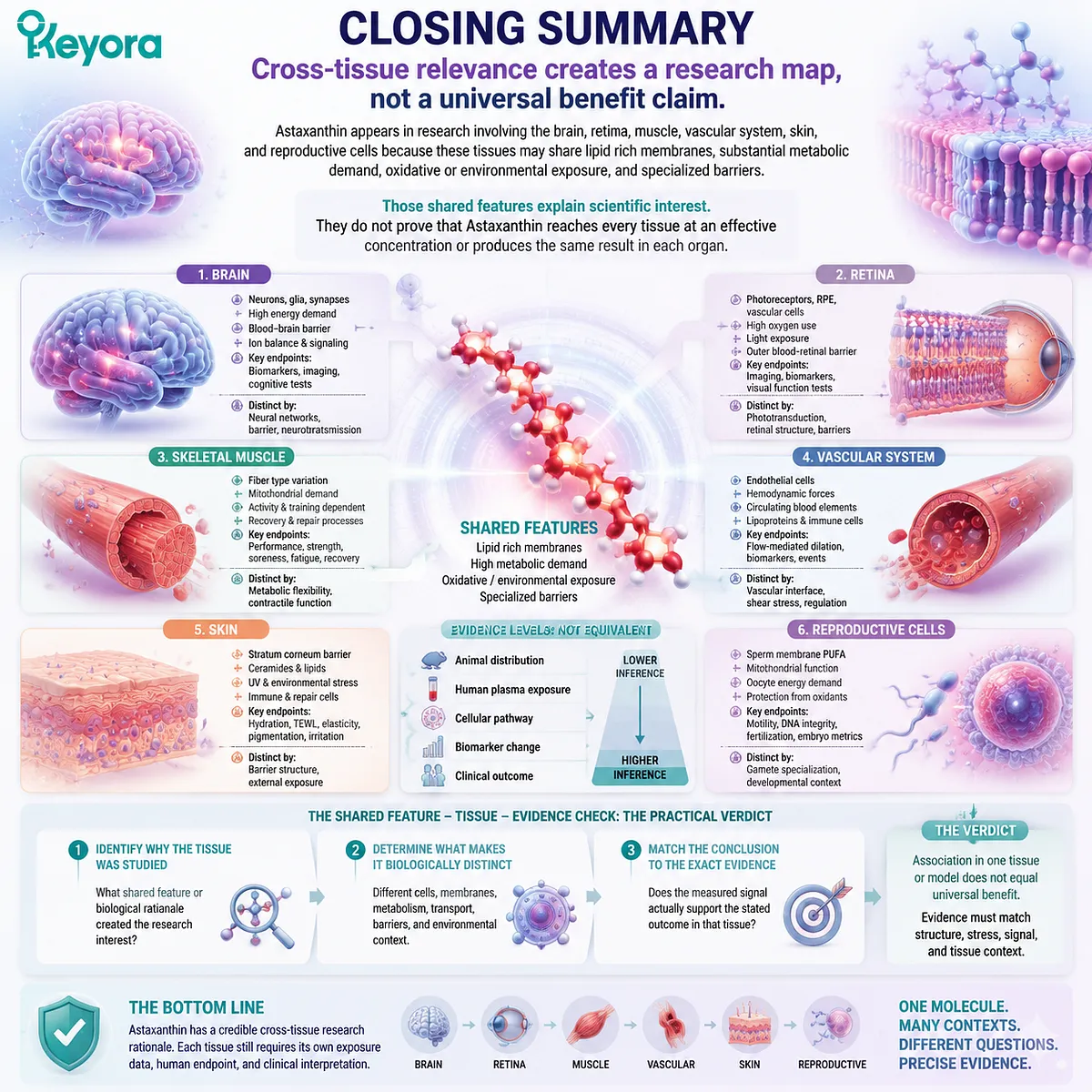
Cross-tissue relevance creates a research map, not a universal benefit claim
Astaxanthin appears in research involving the brain, retina, muscle, vascular system, skin, and reproductive cells because these tissues may share lipid rich membranes, substantial metabolic demand, oxidative or environmental exposure, and specialized barriers.
Those shared features explain scientific interest. They do not prove that Astaxanthin reaches every tissue at an effective concentration or produces the same result in each organ.
Every tissue contains different cells, membranes, metabolic requirements, transport conditions, and meaningful endpoints. Animal distribution, human plasma exposure, a cellular pathway, a biomarker, and a clinical outcome must remain separate evidence levels.
The Shared Feature – Tissue – Evidence Check provides the practical verdict. Identify why the tissue was studied, determine what makes it biologically distinct, and match the conclusion to the exact evidence produced.
Astaxanthin has a credible cross-tissue research rationale. Each tissue still requires its own exposure data, human endpoint, and clinical interpretation.
This article is for educational and informational purposes only. It does not provide medical advice, diagnosis, treatment, cure, prevention, disease outcome claims, hormone restoration claims, fertility outcome claims, or formula-specific clinical efficacy claims.