
How Are Oxidative Stress and Inflammation Connected?
Keyora Research Q&A Library
This is part of the Keyora Research Q&A Series, derived from Keyora Astaxanthin Research Journal Series.
Within the Keyora Astaxanthin Researcn framework, this Q&A translates complex astaxanthin biology into reader-friendly, evidence-bound answers, focusing on natural astaxanthin identity, molecular structure, antioxidant and redox mechanisms, membrane lipid interaction, mitochondrial resilience, inflammatory signaling pathways, human evidence interpretation, and the scientific principles behind responsible supplementation.
First published by Keyora Research Journal: www.keyorahealth.com
Direct Answer
Oxidative stress and inflammation are not the same process, but they can reinforce each other.
Inflammation is an organized immune and tissue response to a trigger such as infection, injury, an allergen, altered self-signals, or metabolic disturbance.
Oxidative stress describes a loss of redox control in which reactive-species production, antioxidant systems, repair, and adaptation are no longer appropriately balanced.
During an inflammatory response, neutrophils, macrophages, and other cells may deliberately produce reactive oxygen species, or ROS. These molecules can help control microorganisms and transmit immune signals.
Human disorders caused by defective phagocyte NADPH oxidase demonstrate that immune ROS have an important host-defense role rather than being accidental waste.
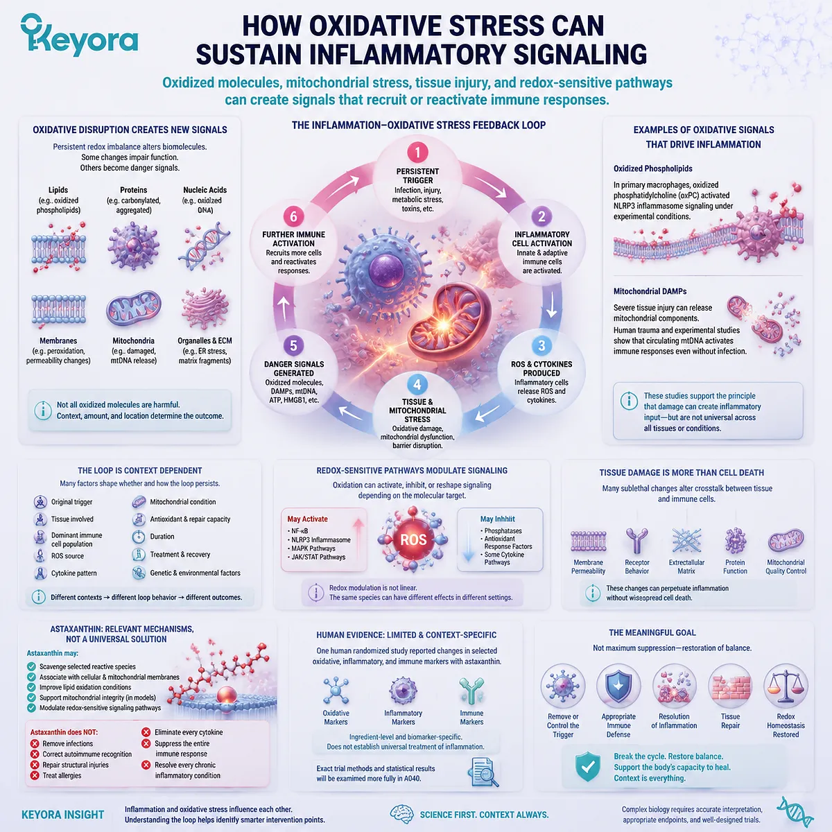
Problems are more likely when the trigger persists or when ROS, cytokines, mitochondrial stress, and tissue damage continue to reinforce one another.
Oxidized lipids and proteins, damaged mitochondria, and injured cells may create new signals that prolong immune activation.
Cell studies have shown that mitochondrial ROS, oxidized phospholipids, and mitochondrial damage signals can activate selected inflammatory systems under defined conditions.
Astaxanthin is relevant because it has been studied in membrane, mitochondrial, redox, and inflammatory-signaling contexts. It is not an anti-inflammatory drug, and antioxidant activity cannot remove every infectious, autoimmune, allergic, or structural trigger.
The practical conclusion is:
Inflammation can increase oxidative pressure, while persistent oxidative disruption can generate signals that sustain inflammation, but neither process explains every case of the other.
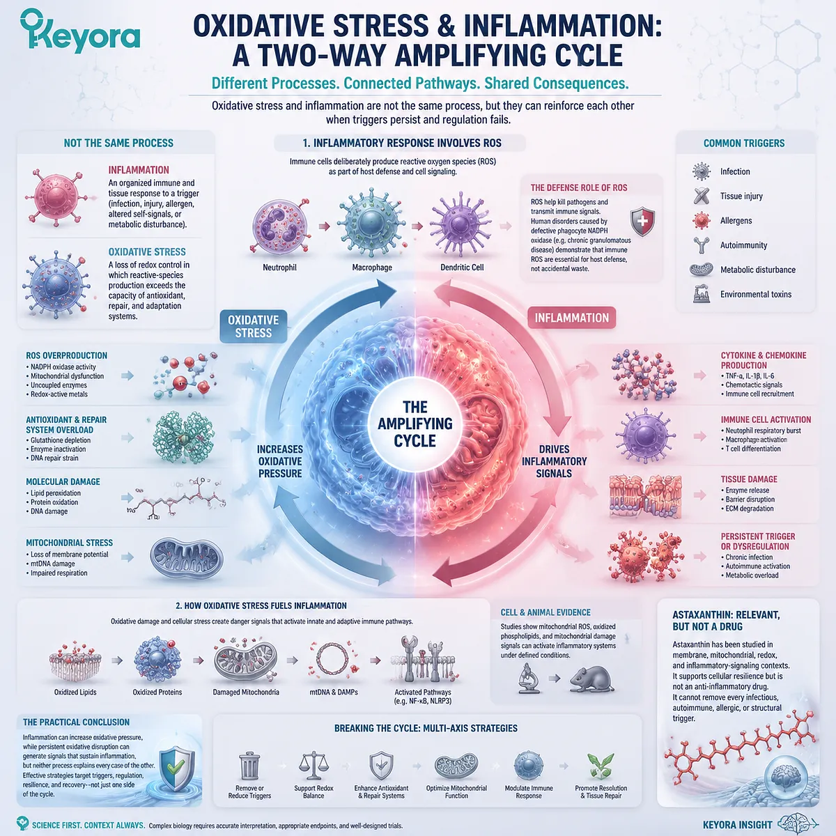
Oxidative Stress and Inflammation Are Different Processes
One describes disrupted redox control, while the other is an organized immune and tissue response to a trigger
Oxidative stress does not mean that any ROS is present. Reactive species are produced during normal metabolism, immune defense, vascular regulation, and adaptation. Oxidative stress becomes the more appropriate description when production, compartmental control, antioxidant processing, repair, and recovery are disturbed enough to alter normal function.
Inflammation has a different definition. It involves coordinated communication among immune cells, blood vessels, tissue cells, signaling molecules, and repair systems in response to a perceived threat or disturbance.
An acute inflammatory response may help:
-
recruit immune cells
-
contain or remove microorganisms
-
clear damaged cells
-
isolate injured tissue
-
initiate repair
-
restore tissue function
Immune ROS are part of this protective biology. Neutrophils activate a nonmitochondrial NADPH oxidase system that transfers electrons to oxygen and supports the respiratory burst. Clinical registries of chronic granulomatous disease, in which NADPH oxidase activity is absent or impaired, show recurrent infection as a major consequence of losing this defense mechanism.
Inflammation therefore should not be described as an error that the body must always suppress. A controlled response can be necessary for survival and healing.
The response also needs to resolve. Resolution involves more than simply reducing one cytokine. The trigger must be controlled or removed, dying cells must be cleared, immune-cell recruitment must decline, and tissue repair must proceed. Experimental studies of macrophage efferocytosis show that clearing apoptotic cells can limit secondary injury and reprogram macrophages toward tissue-repair and resolution functions.
Chronic inflammation is different from a brief protective response. It may persist because of continuing infection, autoimmune activity, repeated injury, barrier disruption, environmental exposure, metabolic disturbance, or failure of normal resolution systems.
The phrase “chronic low-grade inflammation” describes a biological context rather than one specific disease. It cannot be diagnosed from fatigue, weight changes, skin appearance, or discomfort alone.
Oxidative stress and inflammation may occur together, but they can also begin through different routes. An infection can activate inflammation before substantial oxidative damage develops. A chemical or metabolic exposure may disturb redox balance without creating a clinically meaningful inflammatory condition.
The two concepts should therefore be connected without being merged.
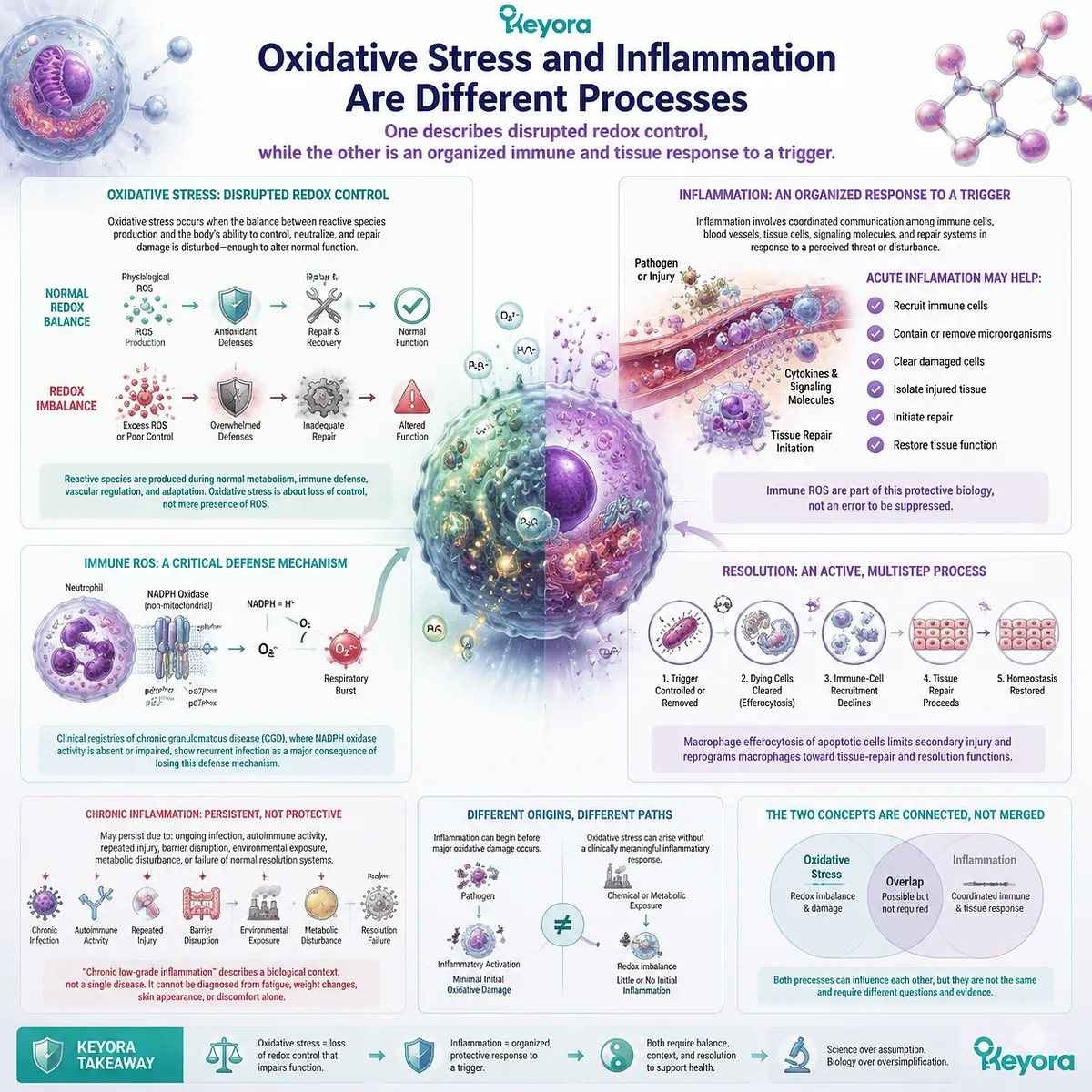
How Inflammation Can Increase Oxidative Pressure
Immune-cell ROS, cytokine signaling, metabolic shifts, and mitochondrial stress can increase reactive-species production during an inflammatory response
When immune cells recognize a microorganism or danger signal, they may increase ROS production rapidly.
In phagocytic cells, the NADPH oxidase respiratory burst is distinct from mitochondrial respiration. Its purpose is not to make ATP. It creates superoxide and downstream reactive chemistry within or near the phagocytic compartment, contributing to antimicrobial defense.
This distinction matters because the phrase “inflammatory ROS” does not identify one source. Reactive species during inflammation may come from:
-
phagocyte NADPH oxidase
-
mitochondria
-
other oxidase enzymes
-
peroxisomes
-
nitric-oxide-related systems
-
extracellular inflammatory chemistry
The biological effect depends on location. ROS generated inside a phagosome to help control a microorganism have a different meaning from persistent oxidant production near healthy membrane proteins or extracellular tissue.
Activated immune and tissue cells may also release cytokines and chemokines. These signals recruit additional cells, alter vascular behavior, change metabolism, and modify cellular redox systems. The response can therefore expand beyond the cell that first detected the trigger.
Redox-sensitive signaling provides another connection. Early primary experiments showed that reactive oxygen intermediates could influence NF-kB activation in selected cell systems. Later experiments also demonstrated that the direction and size of the effect depend on cell type, concentration, timing, and redox-control mechanisms.
NF-kB should not be reduced to an “inflammation switch.” It participates in immunity, cell survival, stress responses, and tissue-specific regulation. A039 will examine its relationship with Astaxanthin and Nrf2 in greater detail.
Inflammatory signaling can also affect mitochondria. Cytokines, altered calcium handling, substrate pressure, and changes in cellular demand may modify mitochondrial metabolism and ROS production. Damaged or poorly controlled mitochondria can then become another contributor to the local inflammatory environment.
A well-known primary cell study found that accumulation of damaged, ROS-generating mitochondria promoted activation of the NLRP3 inflammasome under the tested conditions. This supports one specific mitochondrial inflammation pathway, not a conclusion that all inflammation begins in mitochondria.
The duration of the response is critical.
A short respiratory burst during infection may be protective. Continuous immune-cell activation can expose surrounding lipids, proteins, extracellular structures, and neighboring cells to a different level of oxidative pressure.
The same system can therefore support defense when localized and controlled, yet contribute to collateral tissue stress when activation is excessive, prolonged, or poorly resolved.
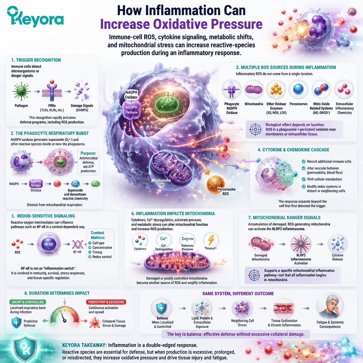
How Oxidative Stress Can Sustain Inflammatory Signaling
Oxidized molecules, mitochondrial stress, tissue injury, and redox-sensitive pathways can create signals that recruit or reactivate immune responses
The feedback loop can also run in the opposite direction.
Persistent oxidative disruption may alter lipids, proteins, nucleic acids, membranes, and organelles. Some altered molecules affect cell function directly. Others become signals that are detected by immune and tissue-surveillance systems.
Oxidized phospholipids provide one example. In a primary macrophage study, oxidized phosphatidylcholine activated NLRP3-related inflammatory signaling under the experimental conditions. The finding supports the principle that an oxidation product can become an inflammatory stimulus, but it does not show that every oxidized lipid activates the same pathway in people.
Mitochondrial damage can also create inflammatory signals.
During severe tissue injury, mitochondrial components may be released from damaged cells. A human trauma and experimental study found that circulating mitochondrial damage-associated molecular patterns activated immune responses, providing evidence that tissue injury can generate new inflammatory input even when infection is not the original trigger.
A simplified feedback route is:
Persistent trigger
→ inflammatory-cell activation
→ ROS and cytokines
→ tissue and mitochondrial stress
→ oxidized or released danger signals
→ further immune activation
This model is useful, but it is not universal.
The loop may differ according to:
-
the original trigger
-
the tissue involved
-
the dominant immune-cell population
-
the ROS source
-
the cytokine pattern
-
mitochondrial condition
-
antioxidant and repair capacity
-
duration
-
treatment and recovery
Tissue damage does not need to mean widespread cell death. Changes in membrane permeability, receptor behavior, extracellular matrix, protein function, and mitochondrial quality control may all alter the signals exchanged between tissue and immune cells.
Redox-sensitive pathways can then influence the intensity or duration of cytokine expression. This does not mean that oxidants always activate inflammation in a simple linear manner. Oxidation can activate, inhibit, or reshape signaling depending on which molecular target is modified.
This context dependence is important for Astaxanthin claims.
Astaxanthin may interact with selected reactive species, associate with membranes, affect lipid-oxidation conditions, and influence redox-sensitive signaling in experimental models. These mechanisms create a credible research rationale for examining the oxidative stress – inflammation loop.
They do not demonstrate that Astaxanthin:
-
removes an infection
-
corrects autoimmune recognition
-
repairs a structural injury
-
treats an allergy
-
eliminates every cytokine
-
suppresses the entire immune response
-
resolves every chronic inflammatory condition
One human randomized study reported changes in selected oxidative, inflammatory, and immune markers after Astaxanthin supplementation. Those results are ingredient-level and biomarker-specific, and they do not establish universal treatment of inflammation. The exact trial methods and statistical results will be examined more fully in A040.
The meaningful goal is not maximum suppression of inflammation. It is removal or control of the trigger, appropriate immune defense, resolution, tissue repair, and restoration of redox homeostasis.
Use the Keyora Trigger – Loop – Outcome Check
Three questions distinguish a measured biological feedback cycle from a broad claim that antioxidants can solve every inflammatory condition
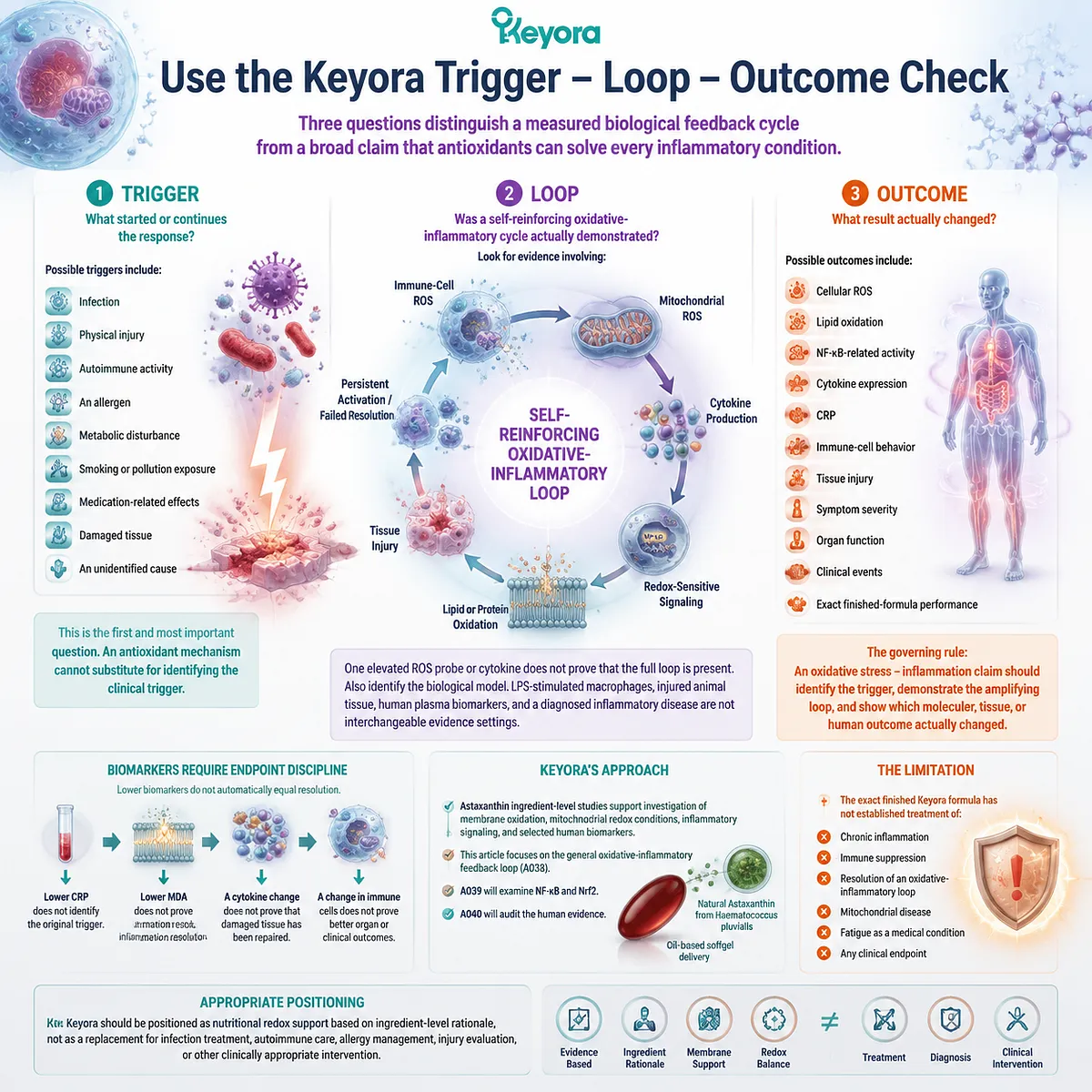
The Keyora Trigger – Loop – Outcome Check provides a practical way to assess claims connecting oxidative stress, inflammation, and Astaxanthin.
1. Trigger
What started or continues the response?
Possible triggers include:
-
infection
-
physical injury
-
autoimmune activity
-
an allergen
-
metabolic disturbance
-
smoking or pollution exposure
-
medication-related effects
-
damaged tissue
-
an unidentified cause
This is the first and most important question. An antioxidant mechanism cannot substitute for identifying the clinical trigger.
2. Loop
Was a self-reinforcing oxidative-inflammatory cycle actually demonstrated?
Look for evidence involving:
-
immune-cell ROS
-
mitochondrial ROS
-
cytokine production
-
redox-sensitive signaling
-
lipid or protein oxidation
-
tissue injury
-
damage-associated signals
-
persistent activation
-
failed resolution
One elevated ROS probe or cytokine does not prove that the full loop is present.
Also identify the biological model. Lipopolysaccharide-stimulated macrophages, injured animal tissue, human plasma biomarkers, and a diagnosed inflammatory disease are not interchangeable evidence settings.
3. Outcome
What result actually changed?
Possible outcomes include:
-
cellular ROS
-
lipid oxidation
-
NF-kB-related activity
-
cytokine expression
-
CRP
-
immune-cell behavior
-
tissue injury
-
symptom severity
-
organ function
-
clinical events
-
exact finished-formula performance
The governing rule is:
An oxidative stress – inflammation claim should identify the trigger, demonstrate the amplifying loop, and show which molecular, tissue, or human outcome actually changed.
Biomarkers also require endpoint discipline. Lower CRP does not identify the original trigger. Lower MDA does not prove inflammation resolution. A cytokine change does not prove that damaged tissue has been repaired.
Astaxanthin is relevant because ingredient-level studies support investigation of membrane oxidation, mitochondrial redox conditions, inflammatory signaling, and selected human biomarkers. The project deliberately separates this general feedback-loop article from A039, which will examine NF-kB and Nrf2, and A040, which will audit the human evidence.
Keyora uses natural Astaxanthin from Haematococcus pluvialis in an oil-based softgel context. This provides a rational formulation setting for a fat-soluble carotenoid, but the exact finished formula has not established treatment of chronic inflammation, immune suppression, or resolution of an oxidative-inflammatory loop.
The product should therefore be positioned as nutritional redox support based on ingredient-level rationale, not as a replacement for infection treatment, autoimmune care, allergy management, injury evaluation, or other clinically appropriate intervention.
Closing Summary
Oxidative stress and inflammation can reinforce each other, but the trigger, duration, control systems, and functional consequences determine their meaning
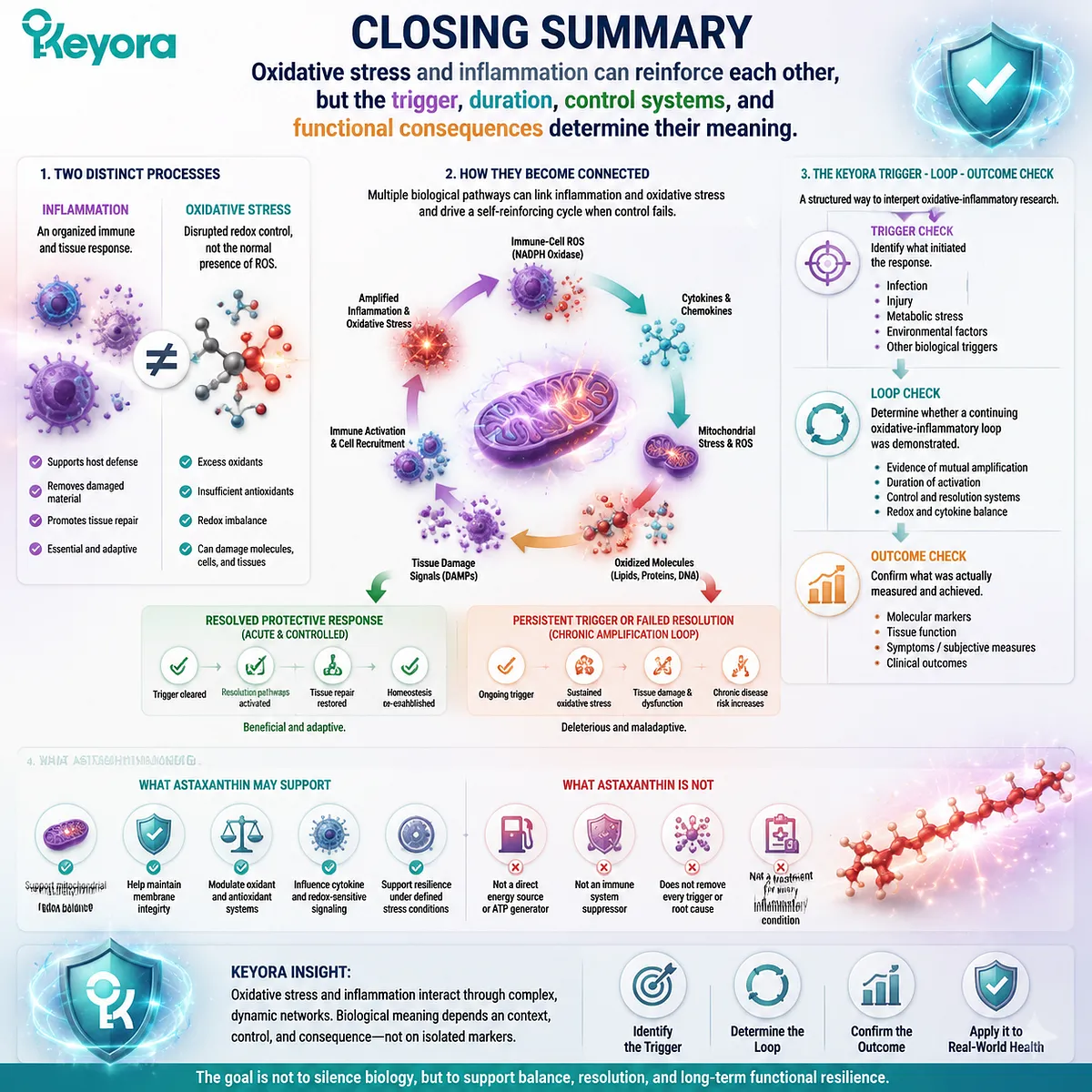
Oxidative stress and inflammation are distinct processes.
Inflammation is an organized immune and tissue response that may support host defense, removal of damaged material, and repair. Oxidative stress describes disrupted redox control rather than the normal presence of ROS.
The two processes become connected when immune-cell ROS, cytokines, mitochondrial stress, oxidized molecules, and tissue-damage signals continue to reinforce one another. A protective acute response may resolve, while a persistent trigger or failed resolution can sustain a chronic amplification loop.
Use the Keyora Trigger – Loop – Outcome Check. Identify what initiated the response, determine whether a continuing oxidative-inflammatory loop was demonstrated, and confirm whether the study measured a molecular marker, tissue function, symptom, or clinical outcome.
Astaxanthin has a credible nutritional role in redox-sensitive inflammatory research. It does not remove every trigger, suppress the entire immune system, or treat every inflammatory condition.
This article is for educational and informational purposes only. It does not provide medical advice, diagnosis, treatment, cure, prevention, disease outcome claims, hormone restoration claims, fertility outcome claims, or formula-specific clinical efficacy claims.