
Does Astaxanthin Support Mitochondrial Function and ATP Production?
Keyora Research Q&A Library
This is part of the Keyora Research Q&A Series, derived from Keyora Astaxanthin Research Journal Series.
Within the Keyora Astaxanthin Researcn framework, this Q&A translates complex astaxanthin biology into reader-friendly, evidence-bound answers, focusing on natural astaxanthin identity, molecular structure, antioxidant and redox mechanisms, membrane lipid interaction, mitochondrial resilience, inflammatory signaling pathways, human evidence interpretation, and the scientific principles behind responsible supplementation.
First published by Keyora Research Journal: www.keyorahealth.com
Direct Answer
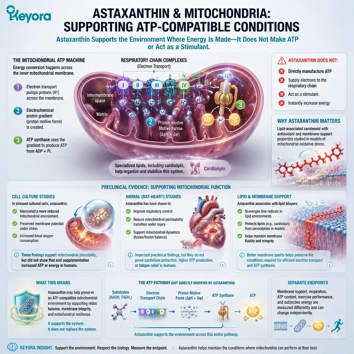
Astaxanthin may support mitochondrial function by helping maintain the membrane and redox conditions in which electron transport and ATP synthesis occur. It does not directly manufacture ATP, supply electrons to the respiratory chain, or act as a stimulant.
The mitochondrial inner membrane contains the respiratory complexes, mobile electron carriers, transport proteins, and ATP synthase.
Electron transport helps create an electrochemical proton gradient across this membrane, while ATP synthase uses that gradient to produce ATP. Specialized lipids, including cardiolipin, contribute to the organization and function of this membrane system.
Astaxanthin is relevant because it is lipid-associated and has been studied in cellular and animal models of mitochondrial oxidative stress.
In cultured cells, astaxanthin helped maintain a more reduced mitochondrial environment, preserved membrane potential under stress, and increased basal oxygen consumption.
These findings support mitochondrial plausibility, but the study did not show that oral supplementation increased ATP production or energy in people.
Rat-heart studies have also reported effects on respiratory control, mitochondrial permeability, and mitochondrial dynamics under experimentally induced injury.
These are important preclinical findings, but they do not prove cardiolipin protection, higher ATP production, or fatigue relief in humans.
The most defensible conclusion is:
Astaxanthin may help preserve an ATP-compatible mitochondrial environment, but membrane support, respiration, ATP content, exercise performance, and subjective energy are separate endpoints.
Why Mitochondrial Membranes Matter for ATP Production
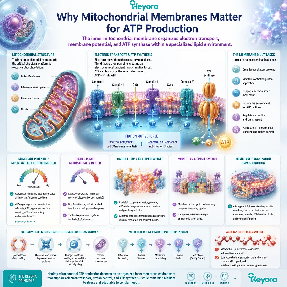
The inner mitochondrial membrane organizes electron transport, membrane potential, and ATP synthase within a specialized lipid environment
Mitochondria contain an outer membrane, an intermembrane space, an inner membrane, and a matrix. The inner mitochondrial membrane is the critical structural platform for oxidative phosphorylation.
Respiratory complexes transfer electrons through a sequence of redox reactions. This transfer is coupled to the movement of protons across the inner membrane, establishing a proton motive force with electrical and concentration components. ATP synthase then uses this stored electrochemical energy to convert ADP and phosphate into ATP.
The membrane must therefore perform several tasks at once:
-
organize respiratory proteins
-
maintain controlled proton separation
-
support electron-carrier movement
-
provide the environment for ATP synthase
-
regulate metabolite and ion transport
-
participate in mitochondrial signaling and quality control
Membrane potential is one part of this system, but it is not identical to ATP production.
A preserved membrane potential can indicate that a mitochondrion retains an important functional condition.
ATP output still depends on substrate availability, ADP supply, oxygen, electron flow, respiratory coupling, ATP synthase activity, and cellular demand.
A higher membrane-potential reading is also not automatically better. Excessive polarization under some metabolic conditions may be associated with restricted electron flow and greater ROS formation, while depolarization can reflect impaired respiratory function or an intentional quality-control response. The meaningful question is whether membrane potential remains appropriately regulated for the biological context.
Cardiolipin is another important part of the inner-membrane environment. It is a distinctive mitochondrial phospholipid associated with respiratory proteins, ATP-related enzymes, membrane curvature, and protein organization. Human and cellular research involving defective cardiolipin remodeling demonstrates that abnormal cardiolipin composition can accompany impaired respiratory and cellular function.
Cardiolipin should not be presented as a single switch controlling mitochondrial energy. The inner membrane also depends on other phospholipids, proteins, membrane-shaping complexes, transporters, respiratory substrates, and mitochondrial quality-control systems.
Primary cell research has shown that altering cardiolipin-associated membrane organization can change respiratory-chain supercomplex formation, membrane potential, ATP-linked respiration, and cell function. This supports the broader principle that mitochondrial energy production depends on organized membrane biology rather than on respiratory enzymes operating in isolation.
Oxidative stress can disrupt this environment through several routes. Persistent lipid oxidation may alter membrane packing. Oxidative modification of respiratory proteins may interfere with electron transfer. Changes in calcium handling or permeability may disturb membrane potential and stress signaling.
These are possible consequences, not an automatic sequence caused by every ROS signal. Mitochondria continuously use antioxidant processing, protein turnover, membrane remodeling, fusion, fission, and mitophagy to maintain function.
Astaxanthin becomes relevant at this level because it is studied as a membrane-associated redox-active carotenoid. The proposed role is support of the environment in which ATP is produced, not direct participation as an energy substrate.
Where Astaxanthin May Support Mitochondrial Resilience
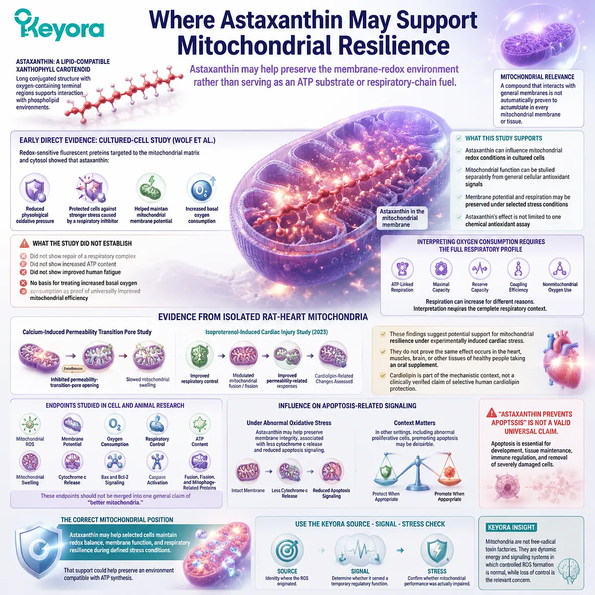
Astaxanthin may help preserve the membrane-redox environment rather than serving as an ATP substrate or respiratory-chain fuel
Astaxanthin has a long lipid-compatible conjugated structure with oxygen-containing terminal regions. Earlier articles in this series established why that architecture supports interaction with phospholipid environments.
Mitochondrial relevance requires an additional step. A compound that interacts with general membranes is not automatically proven to accumulate in every mitochondrial membrane or tissue.
The strongest direct early evidence comes from a cultured-cell study by Wolf and colleagues. The researchers used redox-sensitive fluorescent proteins targeted to the mitochondrial matrix and cytosol. Astaxanthin reduced physiological oxidative pressure and protected cells against stronger stress caused by a respiratory inhibitor. It also helped maintain mitochondrial membrane potential and increased basal oxygen consumption.
These results support several conclusions:
-
astaxanthin can influence mitochondrial redox conditions in cultured cells
-
mitochondrial function can be studied separately from general cellular antioxidant signals
-
membrane potential and respiration may be preserved under selected stress conditions
-
astaxanthin’s effect is not limited to one chemical antioxidant assay
The study did not establish that astaxanthin repaired a respiratory complex, increased ATP content, or improved human fatigue. It also found no basis for treating increased basal oxygen consumption as proof of universally improved mitochondrial efficiency.
Respiration can increase for different reasons. It may reflect greater ATP-linked demand, greater proton leak, altered substrate oxidation, or another shift in cellular metabolism. Interpreting an oxygen-consumption result requires the full respiratory profile, including ATP-linked respiration, maximal capacity, reserve, coupling, and nonmitochondrial oxygen use.
Research in isolated rat-heart mitochondria provides another evidence layer.
One study found that astaxanthin inhibited calcium-induced mitochondrial permeability-transition-pore opening and slowed mitochondrial swelling under the tested conditions.
A later isoproterenol-injury study reported improved respiratory control and changes in mitochondrial fusion, fission, and permeability-related responses after astaxanthin exposure.
These findings suggest potential support for mitochondrial resilience under experimentally induced cardiac stress. They do not prove that the same effect occurs in the heart, muscles, brain, or other tissues of healthy people taking an oral supplement.
The 2023 rat-heart investigation also assessed cardiolipin-related and mitochondrial-dynamics endpoints. That makes cardiolipin part of the mechanistic context, but it does not establish a clinically verified claim that astaxanthin selectively targets or protects human cardiolipin.
Cell and animal studies have additionally examined:
-
mitochondrial ROS
-
membrane potential
-
oxygen consumption
-
respiratory control
-
ATP content
-
mitochondrial swelling
-
cytochrome c release
-
Bax and Bcl-2-related signaling
-
caspase activation
-
fusion, fission, and mitophagy-related proteins
These endpoints should not be merged into one general claim of “better mitochondria.”
Astaxanthin may also influence apoptosis-related signaling under abnormal oxidative stress. In some normal-cell injury models, preservation of membrane integrity has been associated with less cytochrome c release and reduced apoptosis signaling. In other biological settings, including abnormal proliferative cells, promoting apoptosis may be desirable.
This is why “astaxanthin prevents apoptosis” is not a valid universal claim. Apoptosis is an essential biological process involved in development, tissue maintenance, immune regulation, and removal of severely damaged cells.
The correct mitochondrial position is narrower:
Astaxanthin may help selected cells maintain redox balance, membrane function, and respiratory resilience during defined stress conditions.
That support could help preserve an environment compatible with ATP synthesis. It does not mean that astaxanthin molecules are converted into ATP or that every user will feel an energy increase.
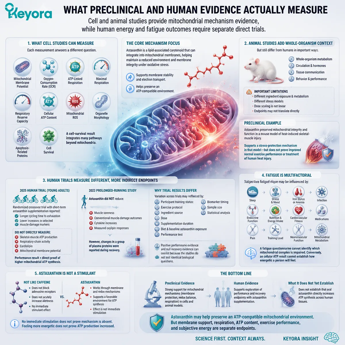
What Preclinical and Human Evidence Actually Measure
Cell and animal studies provide mitochondrial mechanism evidence, while human energy and fatigue outcomes require separate direct trials
The phrase “supports mitochondrial function” can refer to many different measurements.
In cultured cells, investigators can measure:
-
mitochondrial membrane potential
-
oxygen-consumption rate
-
ATP-linked respiration
-
maximal respiration
-
respiratory reserve
-
cellular ATP content
-
mitochondrial ROS
-
organelle morphology
-
apoptosis-related proteins
-
cell survival
Each measurement answers a different question.
Membrane potential shows whether an electrical gradient is maintained. Oxygen consumption measures respiratory activity. ATP content reflects the balance between production and use. A cell-survival result integrates many pathways beyond mitochondria.
Animal studies add whole-organism metabolism, circulation, hormonal responses, tissue communication, and behavior. They still differ from human supplementation in ingredient exposure, metabolism, stress model, dose scaling, and endpoint interpretation.
For example, astaxanthin has preserved mitochondrial integrity and function in a mouse model of heat-induced skeletal-muscle injury. That supports a stress-protection mechanism in that model. It does not establish improved normal exercise performance or treatment of human heat injury.
Human studies usually measure more indirect endpoints.
A 2025 randomized crossover trial in young adults reported longer cycling time to exhaustion and lower increases in selected muscle-damage markers after short-term astaxanthin supplementation.
The study did not directly measure skeletal-muscle ATP production, respiratory-chain activity, cardiolipin, or mitochondrial membrane potential.
The performance result therefore cannot be described as direct proof of higher mitochondrial ATP synthesis.
Other trials have produced different findings. In a 2023 prolonged-running study, astaxanthin did not reduce muscle soreness, conventional muscle-damage outcomes, cytokine increases, or measured oxylipin responses, although changes in a group of plasma proteins were reported during recovery.
Variation across trials may reflect:
-
participant training status
-
exercise protocol
-
ingredient source
-
dose
-
supplementation duration
-
diet
-
baseline astaxanthin exposure
-
performance test
-
biomarker timing
-
sample size
-
statistical analysis
Positive performance evidence and null recovery evidence can coexist because the studies do not test identical biological questions.
Fatigue creates an additional translation problem.
Subjective fatigue may be influenced by:
-
sleep
-
stress and mood
-
iron status or anemia
-
infection
-
endocrine function
-
nutrition and energy intake
-
cardiovascular fitness
-
medications
-
pain
-
training load
-
neuromuscular function
-
mitochondrial metabolism
A fatigue questionnaire cannot identify which mitochondrial complex is impaired. Conversely, a cellular ATP result cannot establish how energetic a person will feel.
Astaxanthin is also not a stimulant. It does not act like caffeine by acutely blocking adenosine receptors and increasing alertness. A lack of immediate stimulation does not prove that a membrane-redox mechanism is absent, while a subjective feeling of energy does not prove that mitochondrial ATP production increased.
The human evidence therefore supports careful exploration of performance and recovery endpoints. It does not yet establish that oral astaxanthin directly increases ATP synthesis across human tissues.
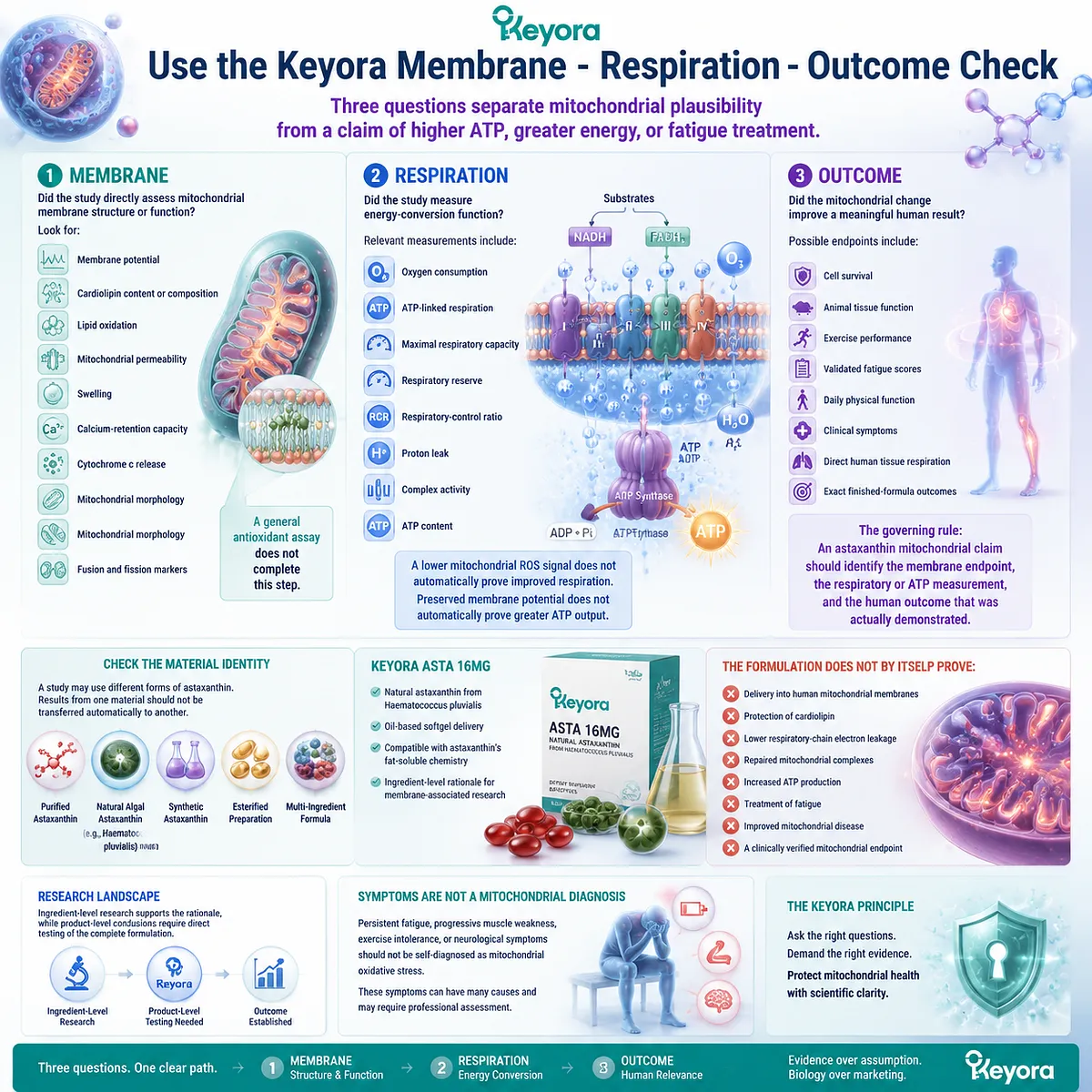
Use the Keyora Membrane – Respiration – Outcome Check
Three questions separate mitochondrial plausibility from a claim of higher ATP, greater energy, or fatigue treatment
The Keyora Membrane – Respiration – Outcome Check provides a practical way to evaluate mitochondrial claims about astaxanthin.
1. Membrane
Did the study directly assess mitochondrial membrane structure or function?
Look for:
-
membrane potential
-
cardiolipin content or composition
-
lipid oxidation
-
mitochondrial permeability
-
swelling
-
calcium-retention capacity
-
cytochrome c release
-
mitochondrial morphology
-
fusion and fission markers
A general antioxidant assay does not complete this step.
2. Respiration
Did the study measure energy-conversion function?
Relevant measurements include:
-
oxygen consumption
-
ATP-linked respiration
-
maximal respiratory capacity
-
respiratory reserve
-
respiratory-control ratio
-
proton leak
-
complex activity
-
ATP content
A lower mitochondrial ROS signal does not automatically prove improved respiration. Preserved membrane potential does not automatically prove greater ATP output.
3. Outcome
Did the mitochondrial change improve a meaningful human result?
Possible endpoints include:
-
cell survival
-
animal tissue function
-
exercise performance
-
validated fatigue scores
-
daily physical function
-
clinical symptoms
-
direct human tissue respiration
-
exact finished-formula outcomes
The governing rule is:
An astaxanthin mitochondrial claim should identify the membrane endpoint, the respiratory or ATP measurement, and the human outcome that was actually demonstrated.
Material identity must also be checked. A study may use purified astaxanthin, natural algal astaxanthin, synthetic astaxanthin, an esterified preparation, or a multi-ingredient formula. Results from one material should not be transferred automatically to another.
Keyora Asta 16MG uses natural astaxanthin from Haematococcus pluvialis in an oil-based softgel context. This formulation is compatible with astaxanthin’s fat-soluble chemistry and provides an ingredient-level rationale for membrane-associated research.
The formulation does not by itself prove:
-
delivery into human mitochondrial membranes
-
protection of cardiolipin
-
lower respiratory-chain electron leakage
-
repaired mitochondrial complexes
-
increased ATP production
-
treatment of fatigue
-
improved mitochondrial disease
-
a clinically verified mitochondrial endpoint
The exact finished Keyora formula has not established a direct mitochondrial, ATP, or fatigue outcome in the supplied evidence corpus. Ingredient-level research supports the rationale, while product-level conclusions require direct testing of the complete formulation.
Persistent fatigue, progressive muscle weakness, exercise intolerance, or neurological symptoms should not be self-diagnosed as mitochondrial oxidative stress. These symptoms can have many causes and may require professional assessment.
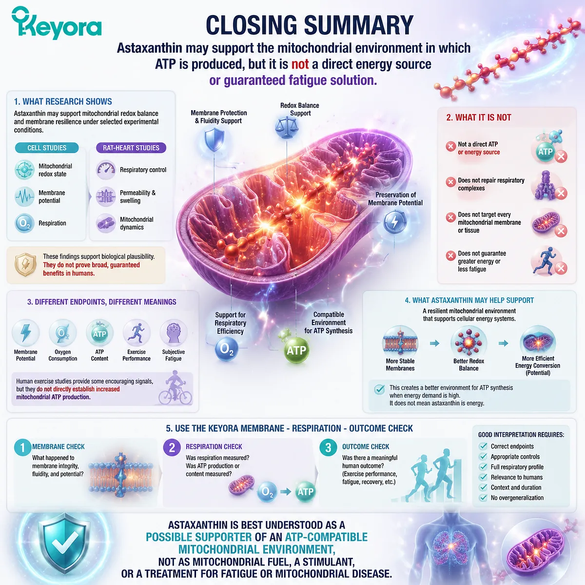
Closing Summary
Astaxanthin may support the mitochondrial environment in which ATP is produced, but it is not a direct energy source or guaranteed fatigue solution
Astaxanthin may support mitochondrial redox balance and membrane resilience under selected experimental conditions. Cell studies report effects on mitochondrial redox state, membrane potential, and respiration, while rat-heart studies provide evidence involving respiratory control, permeability, and mitochondrial dynamics.
These findings support biological plausibility. They do not show that astaxanthin directly creates ATP, repairs respiratory complexes, targets every mitochondrial membrane, or guarantees greater energy.
Membrane potential, oxygen consumption, ATP content, exercise performance, and subjective fatigue are different endpoints. Human exercise studies provide some encouraging signals, but they do not directly establish increased mitochondrial ATP production.
Use the Keyora Membrane – Respiration – Outcome Check. Confirm what happened to the membrane, determine whether respiration or ATP was measured, and identify whether a meaningful human outcome followed.
Astaxanthin is best understood as a possible supporter of an ATP-compatible mitochondrial environment, not as mitochondrial fuel, a stimulant, or a treatment for fatigue or mitochondrial disease.
This article is for educational and informational purposes only. It does not provide medical advice, diagnosis, treatment, cure, prevention, disease outcome claims, hormone restoration claims, fertility outcome claims, or formula-specific clinical efficacy claims.