
Why Do Mitochondria Produce Free Radicals?
Keyora Research Q&A Library
This is part of the Keyora Research Q&A Series, derived from Keyora Astaxanthin Research Journal Series.
Within the Keyora Astaxanthin Researcn framework, this Q&A translates complex astaxanthin biology into reader-friendly, evidence-bound answers, focusing on natural astaxanthin identity, molecular structure, antioxidant and redox mechanisms, membrane lipid interaction, mitochondrial resilience, inflammatory signaling pathways, human evidence interpretation, and the scientific principles behind responsible supplementation.
First published by Keyora Research Journal: www.keyorahealth.com

Direct Answer
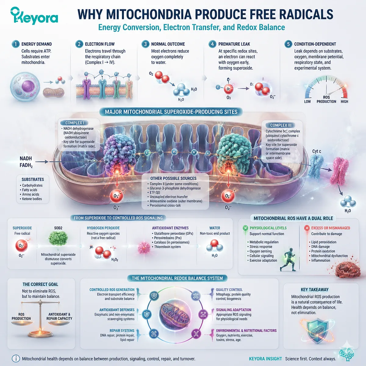
Mitochondria produce free radicals because cellular energy conversion depends on repeated electron-transfer reactions.
Most electrons move through the respiratory chain in a controlled sequence and ultimately contribute to the complete reduction of oxygen to water.
At selected redox sites, however, an electron can react prematurely with oxygen and produce superoxide.
Complex I and Complex III are two important mitochondrial superoxide-producing sites, although they are not the only possible sources. Their contribution changes with nutrient-derived substrates, electron flow, oxygen availability, membrane potential, respiratory state, and the experimental system.
Primary studies using intact mitochondria and isolated respiratory components show that mitochondrial ROS production is highly conditional rather than a fixed percentage of oxygen consumption.
Superoxide is a free radical. Mitochondrial superoxide dismutase helps convert it into hydrogen peroxide, which is a reactive oxygen species but not a free radical. Peroxide-removal systems then regulate its concentration and location.
This chemistry is not automatically evidence of mitochondrial failure. Controlled mitochondrial ROS can participate in metabolic regulation, stress responses, oxygen sensing, and exercise adaptation.
When production becomes persistent, occurs in a damaging location, or exceeds antioxidant, repair, and quality-control capacity, mitochondrial ROS can instead contribute to oxidative stress.
The correct goal is therefore not to eliminate mitochondrial ROS. It is to maintain a system in which ROS generation, signaling, control, repair, and mitochondrial turnover remain appropriately balanced.
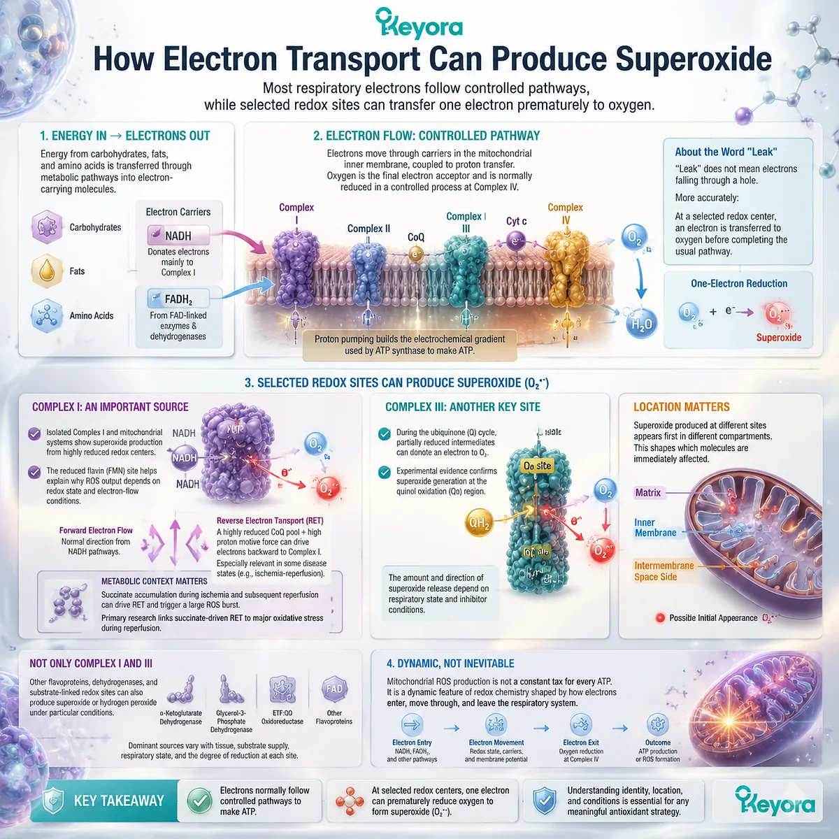
How Electron Transport Can Produce Superoxide
Most respiratory electrons follow controlled pathways, while selected redox sites can transfer one electron prematurely to oxygen
Energy stored in carbohydrates, fats, and amino acids is transferred through metabolic pathways into electron-carrying molecules. NADH supplies electrons mainly to Complex I, while several FAD-linked enzymes and dehydrogenases feed electrons into other parts of the respiratory system.
Electrons then move through carriers in the mitochondrial inner membrane. This movement is coupled to proton transfer and contributes to the electrochemical gradient used by ATP synthase. Oxygen serves as the final electron acceptor and is normally reduced through a controlled process at Complex IV.
The word “leak” is often used when mitochondrial ROS are discussed. It should not be understood as electrons physically falling through a hole in the mitochondrion.
A more accurate description is:
At a selected redox center, an electron is transferred to oxygen before completing the usual respiratory pathway.
The one-electron reduction of oxygen can be represented as:
O₂
-
one electron
→ O₂•−
O₂•− is superoxide.
Complex I is one important source. Experiments with isolated Complex I and mitochondrial systems support superoxide production from highly reduced redox centers within the complex. Research identifying the reduced flavin site also demonstrates why Complex I output depends on its redox state and electron-flow conditions rather than simply on whether the mitochondrion is active.
Complex I can produce ROS during different respiratory states. In some conditions, electrons move in their usual forward direction from NADH-related pathways. In others, a highly reduced ubiquinone pool and a strong proton motive force can support reverse electron transport toward Complex I.
Reverse electron transport is not the normal explanation for every mitochondrial ROS signal. It becomes especially relevant in selected metabolic and pathological settings, including experimental ischemia-reperfusion models. Primary research has linked succinate accumulation and reverse electron flow to a major ROS burst during reperfusion, illustrating how metabolic context can transform the same respiratory machinery into a stronger ROS source.
Complex III is another important site. During the ubiquinone-related Q cycle, partially reduced intermediates can transfer an electron to oxygen under selected conditions. Experimental manipulation of Complex III has confirmed superoxide generation associated with its quinol oxidation region, although the measured amount and direction of release depend on respiratory state and inhibitor conditions.
Location matters. Superoxide produced at different respiratory sites may initially appear in the mitochondrial matrix, the intermembrane-space-facing side, or both, depending on the source. This affects which enzymes, proteins, lipids, and signaling systems encounter the initial reactive species.
Complex I and Complex III should not be described as the only mitochondrial ROS sources. Mitochondria contain additional flavoproteins, dehydrogenases, and substrate-linked redox sites that can produce superoxide or hydrogen peroxide under particular conditions. Comparative work in isolated mitochondria shows that the dominant source changes with tissue, substrate supply, respiratory state, and the degree of reduction at each site.
Mitochondrial ROS production is therefore not a constant tax paid for every ATP molecule. It is a dynamic feature of redox chemistry shaped by how electrons enter, move through, and leave the respiratory system.
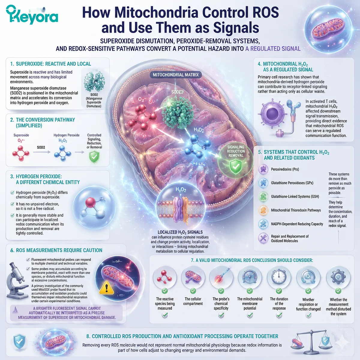
How Mitochondria Control ROS and Use Them as Signals
Superoxide dismutation, peroxide-removal systems, and redox-sensitive pathways convert a potential hazard into a regulated signal
Superoxide is reactive and has limited movement across many biological environments. Manganese superoxide dismutase, or SOD2, is positioned in the mitochondrial matrix and accelerates its conversion into hydrogen peroxide and oxygen.
The simplified reaction is:
Superoxide
→ hydrogen peroxide
→ controlled signaling, reduction, or removal
Hydrogen peroxide differs chemically from superoxide. It has no unpaired electron, so it is not a free radical. It is generally more stable and can participate in localized redox communication when its production and removal are tightly controlled.
Primary cell research has shown that mitochondria-derived hydrogen peroxide can contribute to receptor-linked signaling rather than acting only as cellular waste. In activated T cells, mitochondrial hydrogen peroxide affected downstream signal transmission, providing direct evidence that mitochondrial ROS can serve a regulated communication function.
Mitochondria and surrounding cellular compartments use several systems to control hydrogen peroxide and related oxidants. These include:
-
peroxiredoxins
-
glutathione peroxidases
-
glutathione-linked systems
-
mitochondrial thioredoxin pathways
-
NADPH-dependent reducing capacity
-
repair and replacement of oxidized molecules
These systems do more than remove as much peroxide as possible. They help determine the concentration, duration, and reach of a redox signal.
Hydrogen peroxide can influence selected protein cysteine residues and change protein activity, localization, or interactions. This creates a mechanism through which mitochondrial metabolism can communicate with enzymes, transcriptional responses, stress pathways, and other cellular systems.
The location of production remains critical. A small, temporary peroxide signal near a responsive protein may support regulation. The same total amount generated continuously in a poorly controlled region may produce a different result.
ROS measurements also require caution. Fluorescent mitochondrial probes can respond to multiple chemical and technical variables. Some probes may accumulate according to membrane potential, react with more than one species, or disturb mitochondrial function when used at excessive concentrations.
A primary investigation of the commonly used MitoSOX probe found that its accumulation and oxidation products could themselves impair mitochondrial respiration under certain experimental conditions. This shows why a brighter fluorescent signal cannot automatically be interpreted as a precise measurement of superoxide or mitochondrial damage.
A valid mitochondrial ROS conclusion should therefore consider:
-
the reactive species being measured
-
the cellular compartment
-
the probe’s chemical specificity
-
the mitochondrial membrane potential
-
the duration of the response
-
whether respiration or function changed
-
whether the measurement method disturbed the system
Controlled ROS production and antioxidant processing operate together. Removing every ROS molecule would not represent normal mitochondrial physiology because redox information is part of how cells adjust to changing energy and environmental demands.
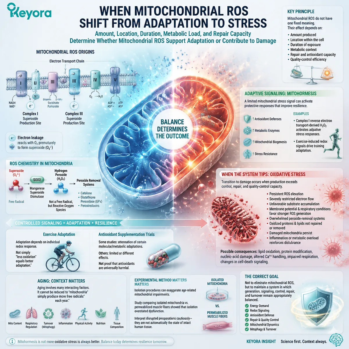
When Mitochondrial ROS Shift From Adaptation to Stress
Amount, location, duration, metabolic load, and repair capacity determine whether mitochondrial ROS support adaptation or contribute to damage
Mitochondrial ROS do not have one fixed biological meaning. Their effect depends on how much is produced, where it appears, how long it persists, and whether antioxidant, repair, and quality-control systems respond successfully.
A temporary increase can function as an adaptive signal. This principle is often described as mitohormesis.
Mitohormesis means that a limited mitochondrial stress signal can activate responses that improve resilience to a later challenge.
It does not mean that more oxidative stress is always better.
Primary experimental work has shown that hydrogen peroxide derived from Complex I during reverse electron transport can activate adaptive stress responses. In that model, suppressing the signal reduced survival under subsequent stress, demonstrating that selected mitochondrial ROS can carry useful biological information.
Exercise provides a practical example. Muscle contraction changes energy demand, oxygen use, redox state, calcium handling, and ROS production. The resulting redox signals can contribute to changes in antioxidant defenses, metabolic enzymes, and endurance-related adaptation.
A controlled exercise study found that training adaptation depended partly on the participants’ exercise-induced redox response. The relationship was not simply “less oxidation equals better adaptation.” Individuals with different redox responses showed different training outcomes, supporting a context-dependent model.
Other human trials have examined whether high-dose antioxidant supplementation changes training responses. Some reported attenuation of selected molecular or metabolic adaptations, while others found more limited or different effects. These results do not prove that antioxidants are universally harmful during exercise. They show that completely suppressing redox signals is not necessarily the same as supporting adaptation.
The transition from adaptation to oxidative stress can occur when:
-
ROS production remains elevated
-
electron flow becomes severely restricted
-
substrates accumulate in an unfavorable redox state
-
membrane potential and respiratory conditions favor stronger ROS generation
-
peroxide-removal systems are overwhelmed
-
oxidized proteins and lipids are not repaired or removed
-
damaged mitochondria persist
-
inflammation or metabolic overload reinforces the disturbance
Under those conditions, mitochondrial ROS may contribute to lipid oxidation, protein modification, nucleic-acid damage, altered calcium handling, impaired respiratory function, and changes in cell-death signaling. The presence of one ROS signal alone does not establish that all these outcomes occurred.
Aging should also be interpreted carefully. Aging can be accompanied by changes in mitochondrial content, respiratory regulation, turnover, inflammation, physical activity, and tissue composition. It cannot be reduced to one claim that mitochondria simply produce more free radicals each year.
Experimental method can strongly influence the conclusion. A study comparing isolated mitochondria with permeabilized muscle fibers found that isolation procedures exaggerated some apparent age-related mitochondrial impairments. This demonstrates why results from disrupted preparations must not be treated automatically as the state of intact human tissue.
Healthy mitochondrial aging therefore depends on more than radical removal. It also involves energy demand, mitochondrial biogenesis, fission and fusion, mitophagy, protein quality control, repair, physical activity, nutrition, and the broader inflammatory environment.
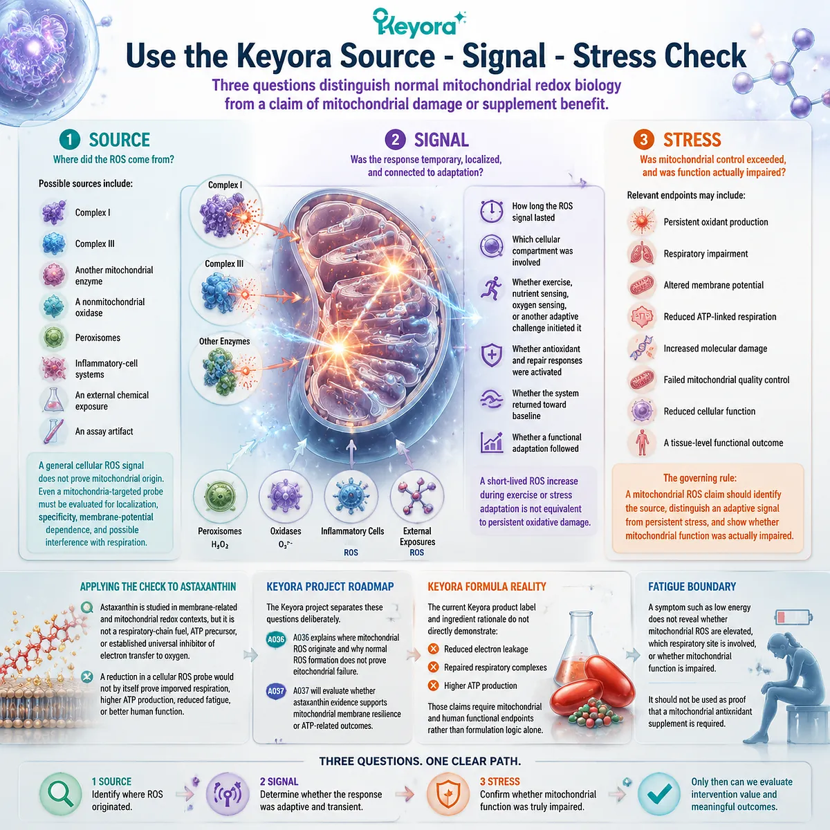
Use the Keyora Source – Signal – Stress Check
Three questions distinguish normal mitochondrial redox biology from a claim of mitochondrial damage or supplement benefit
The Keyora Source – Signal – Stress Check provides a practical way to evaluate statements about mitochondrial free radicals.
1. Source
Where did the ROS come from?
Possible sources include:
-
Complex I
-
Complex III
-
another mitochondrial enzyme
-
a nonmitochondrial oxidase
-
peroxisomes
-
inflammatory-cell systems
-
an external chemical exposure
-
an assay artifact
A general cellular ROS signal does not prove mitochondrial origin. Even a mitochondria-targeted probe must be evaluated for localization, specificity, membrane-potential dependence, and possible interference with respiration.
2. Signal
Was the response temporary, localized, and connected to adaptation?
Check:
-
how long the ROS signal lasted
-
which cellular compartment was involved
-
whether exercise, nutrient sensing, oxygen sensing, or another adaptive challenge initiated it
-
whether antioxidant and repair responses were activated
-
whether the system returned toward baseline
-
whether a functional adaptation followed
A short-lived ROS increase during exercise or stress adaptation is not equivalent to persistent oxidative damage.
3. Stress
Was mitochondrial control exceeded, and was function actually impaired?
Relevant endpoints may include:
-
persistent oxidant production
-
respiratory impairment
-
altered membrane potential
-
reduced ATP-linked respiration
-
increased molecular damage
-
failed mitochondrial quality control
-
reduced cellular function
-
a tissue-level functional outcome
The governing rule is:
A mitochondrial ROS claim should identify the source, distinguish an adaptive signal from persistent stress, and show whether mitochondrial function was actually impaired.
This rule is also necessary before evaluating astaxanthin.
Astaxanthin is studied in membrane-related and mitochondrial redox contexts, but it is not a respiratory-chain fuel, ATP precursor, or established universal inhibitor of electron transfer to oxygen. A reduction in a cellular ROS probe would not by itself prove improved respiration, higher ATP production, reduced fatigue, or better human function.
The Keyora project separates these questions deliberately. A036 explains where mitochondrial ROS originate and why normal ROS formation does not prove mitochondrial failure. A037 will evaluate whether astaxanthin evidence supports mitochondrial membrane resilience or ATP-related outcomes.
The current Keyora product label and ingredient rationale do not directly demonstrate reduced electron leakage, repaired respiratory complexes, or higher ATP production for the finished formula. Those claims require mitochondrial and human functional endpoints rather than formulation logic alone.
Fatigue provides an important boundary. A symptom such as low energy does not reveal whether mitochondrial ROS are elevated, which respiratory site is involved, or whether mitochondrial function is impaired. It should not be used as proof that a mitochondrial antioxidant supplement is required.
Closing Summary
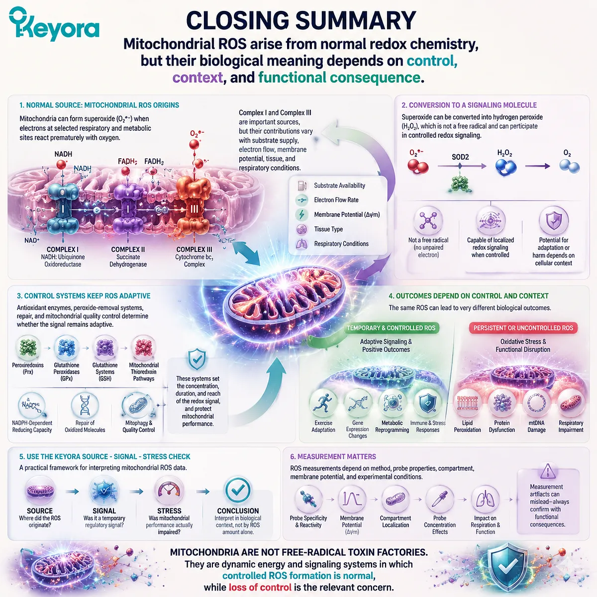
Mitochondrial ROS arise from normal redox chemistry, but their biological meaning depends on control, context, and functional consequence
Mitochondria can form superoxide when electrons at selected respiratory and metabolic sites react prematurely with oxygen. Complex I and Complex III are important sources, but their contributions vary with substrate supply, electron flow, membrane potential, tissue, and respiratory conditions.
Superoxide can be converted into hydrogen peroxide, which is not a free radical and can participate in controlled redox signaling. Antioxidant enzymes, peroxide-removal systems, repair, and mitochondrial quality control determine whether that signal remains adaptive.
Temporary mitochondrial ROS can contribute to exercise responses and other forms of cellular adaptation. Persistent or poorly controlled production can instead contribute to oxidative stress and functional disruption.
Use the Keyora Source – Signal – Stress Check. Identify where the ROS originated, determine whether it served a temporary regulatory function, and confirm whether mitochondrial performance was actually impaired.
Mitochondria are not free-radical toxin factories. They are dynamic energy and signaling systems in which controlled ROS formation is normal, while loss of control is the relevant concern.
This article is for educational and informational purposes only. It does not provide medical advice, diagnosis, treatment, cure, prevention, disease outcome claims, hormone restoration claims, fertility outcome claims, or formula-specific clinical efficacy claims.