
What Makes the Molecular Structure of Astaxanthin Different From Other Carotenoids?
Keyora Research Q&A Library
This is part of the Keyora Research Q&A Series, derived from Keyora Astaxanthin Research Journal Series.
Within the Keyora Astaxanthin Researcn framework, this Q&A translates complex astaxanthin biology into reader-friendly, evidence-bound answers, focusing on natural astaxanthin identity, molecular structure, antioxidant and redox mechanisms, membrane lipid interaction, mitochondrial resilience, inflammatory signaling pathways, human evidence interpretation, and the scientific principles behind responsible supplementation.
First published by Keyora Research Journal: www.keyorahealth.com
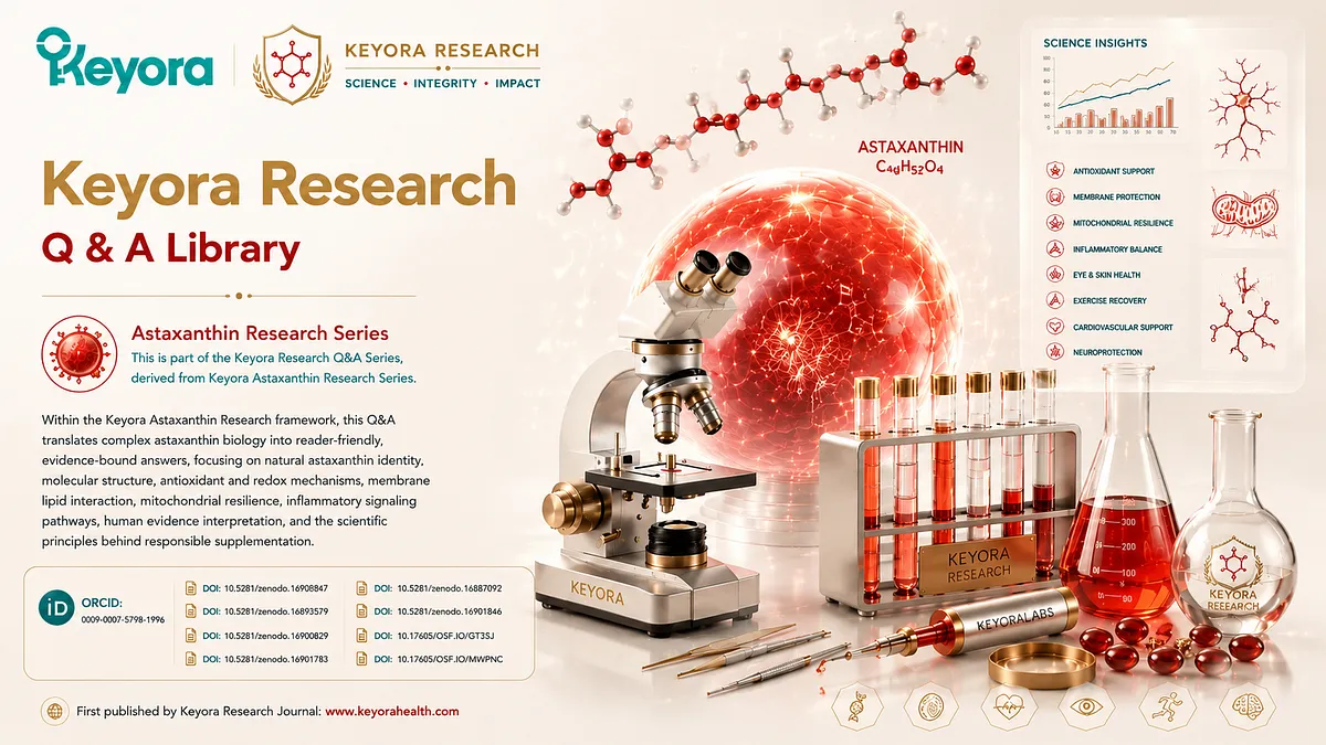
Direct Answer
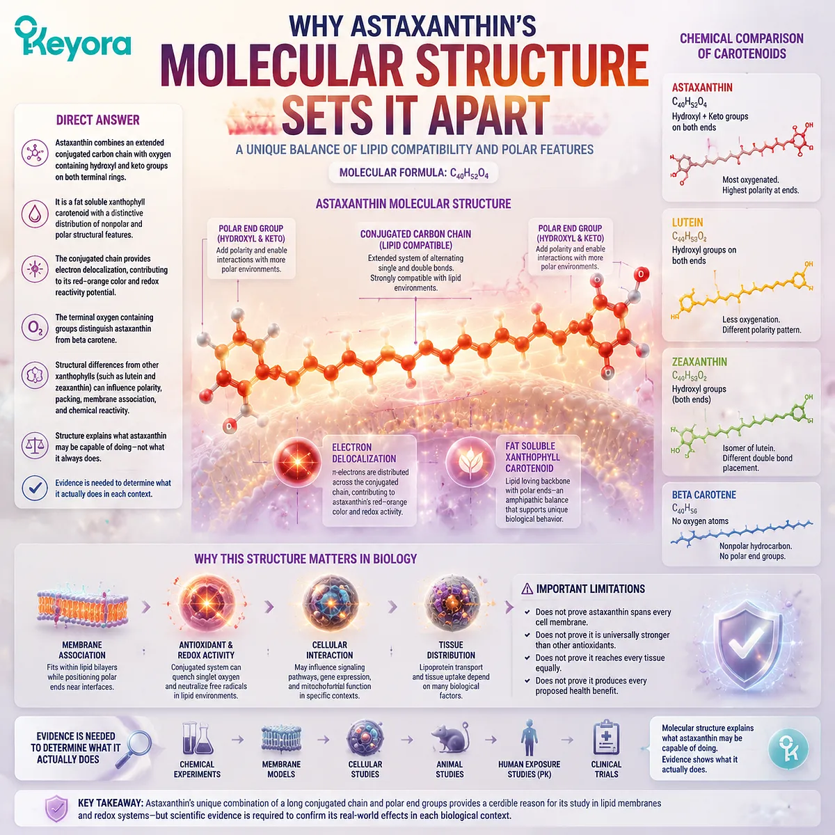
Astaxanthin differs from many other carotenoids because it combines an extended conjugated carbon chain with oxygen containing hydroxyl and keto groups on both terminal rings. The central chain is strongly compatible with lipid environments, while the terminal groups create regions with greater polarity.
Astaxanthin is therefore a fat soluble xanthophyll carotenoid with a distinctive distribution of nonpolar and polar structural features. Its molecular formula is C40H52O4.
The conjugated chain contains alternating single and double bonds. This allows electrons to be distributed across a large part of the molecule, contributing to astaxanthin’s red orange color and its ability to participate in selected reactions involving radicals and electronically excited species.
Its terminal oxygen containing groups distinguish astaxanthin from beta carotene, which contains no oxygen atoms.
Lutein and zeaxanthin are also xanthophyll carotenoids, but their oxygenation patterns differ because they contain hydroxyl groups without astaxanthin’s additional keto groups. These structural differences can influence polarity, molecular packing, membrane association, and chemical reactivity.
Astaxanthin’s structure provides a credible reason to study it in lipid membranes and redox related systems. It does not prove that astaxanthin always spans every cell membrane, is universally stronger than other antioxidants, reaches every tissue, or produces every proposed health benefit.
Molecular structure explains what astaxanthin may be capable of doing.
Chemical experiments, membrane models, human exposure studies, and clinical trials are still needed to determine what it actually does in each biological context.
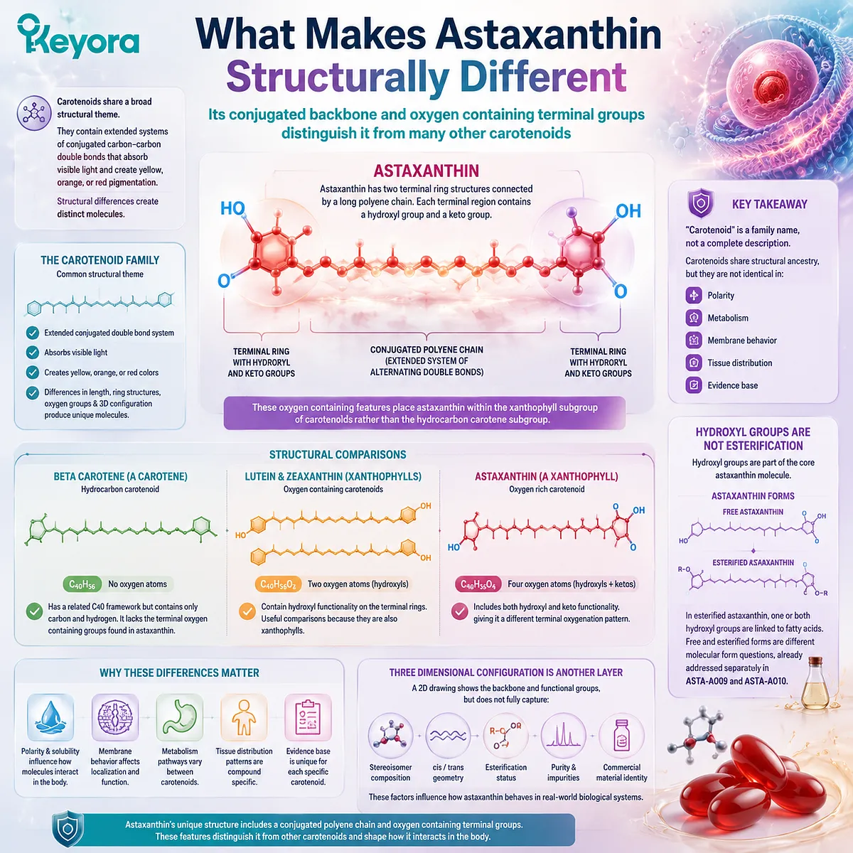
What Makes Astaxanthin Structurally Different
Its conjugated backbone and oxygen containing terminal groups distinguish it from many other carotenoids
Carotenoids share a broad structural theme. They contain extended systems of conjugated carbon to carbon double bonds that absorb visible light and create yellow, orange, or red pigmentation. Differences in the length and arrangement of this conjugated system, terminal rings, oxygen containing groups, and three dimensional configuration produce distinct carotenoid molecules.
Astaxanthin has two terminal ring structures connected by a long polyene chain. Each terminal region contains a hydroxyl group and a keto group. These oxygen containing features place astaxanthin within the xanthophyll subgroup of carotenoids rather than the hydrocarbon carotene subgroup.
Beta carotene provides a useful comparison. It has a related C40 carotenoid framework but contains only carbon and hydrogen. It lacks the terminal oxygen containing groups found in astaxanthin. This difference affects polarity and molecular interactions, but it does not establish a simple hierarchy in which one carotenoid is universally better than another.
Lutein and zeaxanthin are closer comparisons because they are also oxygen containing xanthophylls. Both have the molecular formula C40H56O2 and contain hydroxyl functionality. Astaxanthin has the molecular formula C40H52O4 and includes both hydroxyl and keto functionality, giving it a different terminal oxygenation pattern.
These comparisons show that “carotenoid” is a family name rather than a complete description of molecular behavior. Beta carotene, lutein, zeaxanthin, and astaxanthin share structural ancestry, but they are not identical in polarity, metabolism, membrane behavior, tissue distribution, or evidence base.
Astaxanthin’s hydroxyl groups should also not be confused with esterification. Hydroxyl groups are part of the core astaxanthin molecule. In esterified astaxanthin, one or both hydroxyl groups are linked to fatty acids. Free and esterified forms are therefore different molecular form questions, already addressed separately in ASTA-A009 and ASTA-A010.
Three dimensional configuration is another separate layer. A two dimensional drawing can show the common astaxanthin backbone and functional groups, but it does not fully represent stereoisomer composition, cis and trans geometry, esterification, purity, or commercial material identity.
Why the Conjugated Chain Matters
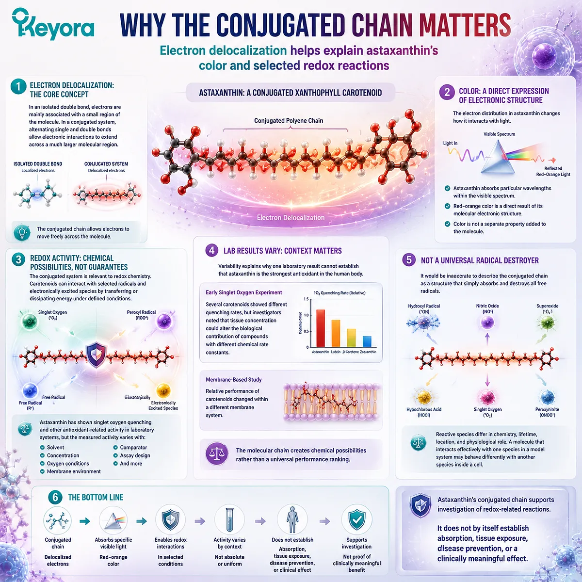
Electron delocalization helps explain astaxanthin’s color and selected redox reactions
In an isolated double bond, electrons are mainly associated with a small region of the molecule. In a conjugated system, alternating single and double bonds allow electronic interactions to extend across a much larger molecular region.
This electron distribution changes how the molecule interacts with light. Astaxanthin absorbs particular wavelengths within the visible spectrum and reflects the red orange color associated with the pigment. The color is therefore a direct expression of molecular electronic structure, not a separate property added to the molecule.
The conjugated system is also relevant to redox chemistry. Carotenoids can interact with selected radicals and electronically excited species by transferring or dissipating energy under defined conditions. Astaxanthin has shown singlet oxygen quenching and other antioxidant related activity in laboratory systems, but the measured activity varies with the solvent, concentration, oxygen conditions, membrane environment, comparator, and assay design.
This variability explains why one laboratory result cannot establish that astaxanthin is the strongest antioxidant in the human body. In one early singlet oxygen experiment, several carotenoids showed different quenching rates, but the investigators also noted that tissue concentration could alter the biological contribution of compounds with different chemical rate constants. Another study found that the relative performance of carotenoids changed within a different membrane based system.
The molecular chain therefore creates chemical possibilities rather than a universal performance ranking.
It would also be inaccurate to describe the conjugated chain as a structure that simply absorbs and destroys all free radicals. Reactive species differ in chemistry, lifetime, location, and physiological role. A molecule that interacts effectively with one species in a model system may behave differently with another species inside a cell.
Astaxanthin’s conjugated chain supports investigation of redox related reactions. It does not by itself establish absorption, tissue exposure, disease prevention, or a clinically meaningful effect.
Why Astaxanthin Is Lipid Associated but Partly Polar
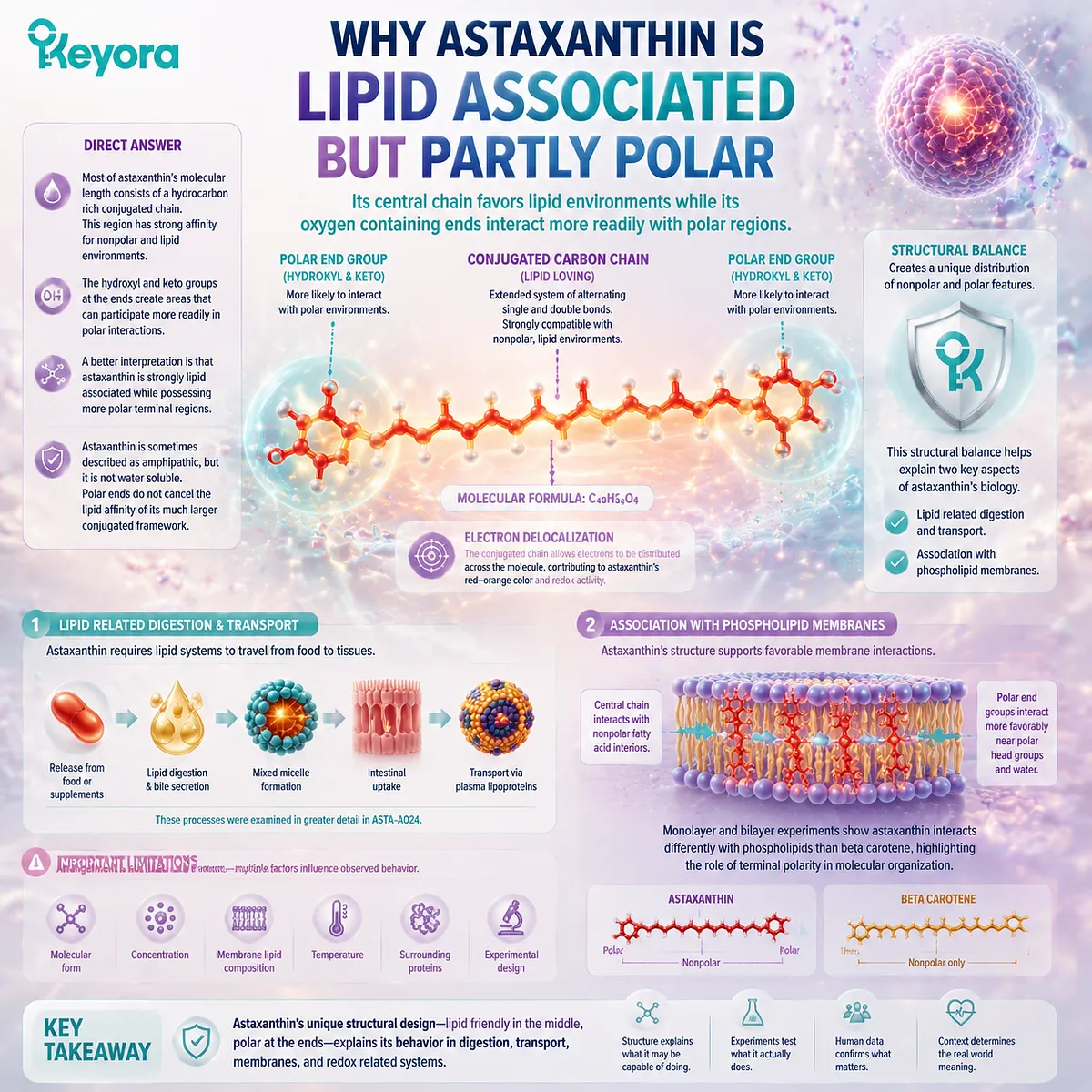
Its central chain favors lipid environments while its oxygen containing ends interact more readily with polar regions
Most of astaxanthin’s molecular length consists of a hydrocarbon rich conjugated chain. This region has strong affinity for nonpolar and lipid environments. The hydroxyl and keto groups at the ends create areas that can participate more readily in polar interactions.
Astaxanthin is sometimes described as amphipathic because it combines these different structural regions. That description requires care. Astaxanthin does not behave like a freely water soluble compound, and the presence of polar end groups does not cancel the lipid affinity of its much larger conjugated framework.
A better interpretation is that astaxanthin is strongly lipid associated while possessing more polar terminal regions.
This structural balance helps explain two different aspects of its biology.
First, astaxanthin requires lipid related digestion and transport. Its fat soluble character influences release from food or supplements, incorporation into mixed micelles, intestinal uptake, and transport through plasma lipoproteins. Those processes were examined in greater detail in ASTA-A024.
Second, the distribution of polarity across the molecule creates a plausible basis for association with phospholipid membranes. A membrane contains a nonpolar interior formed by fatty acid chains and more polar interfaces formed by phospholipid head groups and surrounding water.
Astaxanthin’s central region can interact with lipid chains, while its oxygen containing ends can interact more favorably near polar membrane regions. Monolayer and bilayer experiments have found that astaxanthin interacts differently with phospholipids than beta carotene, supporting the view that terminal polarity affects molecular organization within lipid systems.
This does not mean that astaxanthin has one rigid position under every condition. Molecular form, concentration, membrane lipid composition, temperature, surrounding proteins, and experimental design can all influence the observed arrangement.
What the Structure Suggests About Membranes and Redox Biology
Molecular architecture supports membrane and redox hypotheses without proving one universal orientation or outcome
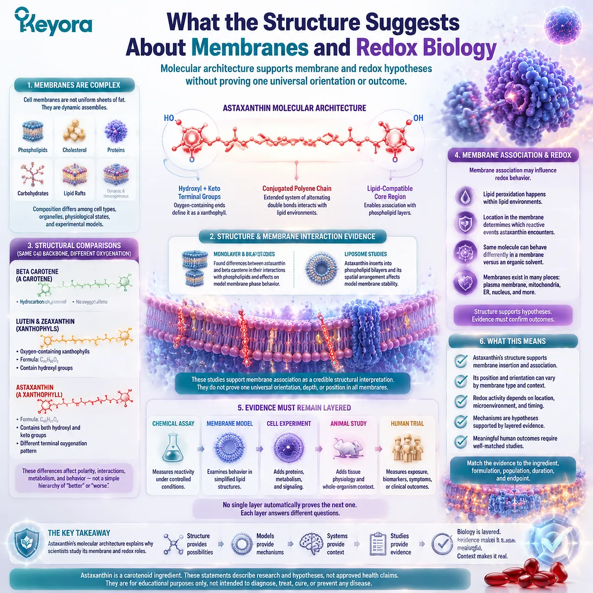
Cell membranes are not uniform sheets of fat. They are dynamic assemblies containing phospholipids, cholesterol, proteins, carbohydrates, and specialized lipid regions. Their composition differs among cell types, organelles, physiological states, and experimental models.
Because astaxanthin contains a long lipid compatible region and oxygen containing ends, researchers have investigated whether it can insert into phospholipid layers and influence membrane organization. A primary monolayer and bilayer study found differences between astaxanthin and beta carotene in their interactions with phospholipids and their effects on model membrane phase behavior.
A later liposome study also reported that astaxanthin inserted into phospholipid bilayers and that its spatial arrangement affected model membrane stability. Liposomes are useful experimental systems, but they are simplified structures and are not equivalent to every membrane in the human body.
These studies support membrane association as a credible structural interpretation. They do not establish that every astaxanthin molecule always spans the complete bilayer, anchors at an identical angle, or occupies the same position in all cell and mitochondrial membranes.
Membrane association may also influence redox behavior. Lipid peroxidation develops within lipid environments, so the location of a carotenoid can affect which reactive events it encounters. A molecule positioned near oxidation sensitive membrane lipids may behave differently from the same molecule dispersed in an organic solvent during a chemical assay.
This is why the evidence must remain layered:
-
A chemical assay measures reactivity under controlled conditions
-
A membrane model examines behavior within a simplified lipid structure
-
A cell experiment adds proteins, metabolism, and signaling
-
An animal study adds tissue physiology
-
A human trial measures exposure, biomarkers, symptoms, or clinical outcomes
No single layer automatically proves the next one.
Astaxanthin’s structure supports hypotheses involving membrane lipids, excited state chemistry, radicals, and redox sensitive signaling. Whether those mechanisms produce a meaningful human outcome must be evaluated with evidence matched to the ingredient, formulation, population, duration, and endpoint.
Use the Keyora Backbone – End Groups – Evidence Check
Three questions help translate a molecular diagram without turning structure into marketing proof
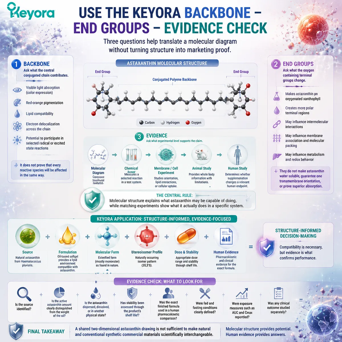
The Keyora Backbone – End Groups – Evidence Check provides a practical way to interpret claims about astaxanthin’s molecular structure.
1. Backbone
Ask what the central conjugated chain contributes.
The chain helps explain visible light absorption, red orange pigmentation, lipid compatibility, electron distribution, and the potential to participate in selected radical or excited state reactions. It does not prove that every reactive species will be affected in the same way.
2. End Groups
Ask what the oxygen containing terminal groups change.
The hydroxyl and keto groups make astaxanthin an oxygenated xanthophyll and create more polar terminal regions. They may influence intermolecular interactions, membrane association, molecular packing, metabolism, and redox behavior.
The end groups do not make astaxanthin water soluble, guarantee one transmembrane orientation, or prove superior absorption.
3. Evidence
Ask what experimental level supports the claim.
A molecular diagram can confirm structural features. A chemical assay can measure a selected reaction. A membrane experiment can study orientation or lipid interactions. Only human research can determine whether supplementation changes a relevant human endpoint.
The central rule is:
Molecular structure explains what astaxanthin may be capable of doing, while matching experiments show what it actually does in a specific system.
Keyora uses natural astaxanthin from Haematococcus pluvialis in an oil based softgel context. The lipid environment is compatible with astaxanthin’s lipid associated structure, but structural compatibility does not prove superior absorption, a fixed membrane orientation, or a clinical outcome for the exact finished formula.
The commercial material must still be matched by source, molecular form, stereoisomer profile, formulation, dose, and evidence. A shared two dimensional astaxanthin drawing is not sufficient to make natural and conventional synthetic commercial materials scientifically interchangeable.
Closing Summary
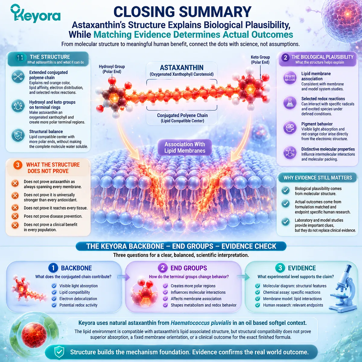
Astaxanthin’s structure explains biological plausibility, while matching evidence determines actual outcomes
Astaxanthin combines an extended conjugated polyene chain with hydroxyl and keto groups on its terminal rings. The chain helps explain its red orange color, lipid affinity, electron distribution, and selected redox reactions. The oxygen containing ends make astaxanthin a xanthophyll and create more polar terminal regions without making the complete molecule water soluble.
This structural balance provides a credible reason to study astaxanthin in lipid membranes and redox related systems. Membrane models support lipid bilayer association, while chemical studies show that astaxanthin can interact with selected radicals and excited species under defined conditions.
These findings do not prove that astaxanthin always spans every membrane, is universally stronger than every antioxidant, reaches every tissue, prevents disease, or produces a clinical benefit in every population.
Use the Keyora Backbone – End Groups – Evidence Check. Identify what the conjugated chain contributes, examine how the terminal groups change molecular behavior, and then confirm which evidence level supports the proposed effect.
Astaxanthin’s structure establishes a mechanism foundation. It does not replace formulation matched and endpoint specific human evidence.
This article is for educational and informational purposes only. It does not provide medical advice, diagnosis, treatment, cure, prevention, disease outcome claims, hormone restoration claims, fertility outcome claims, or formula-specific clinical efficacy claims.